. 2009 Aug 27:10:403.
doi: 10.1186/1471-2164-10-403.

Transcriptional profiling reveals developmental relationship and distinct biological functions of CD16+ and CD16- monocyte subsets

Affiliations

Transcriptional profiling reveals developmental relationship and distinct biological functions of CD16+ and CD16- monocyte subsets

Petronela Ancuta et al. BMC Genomics. .

Abstract

Background: Human peripheral blood monocytes (Mo) consist of subsets distinguished by expression of CD16 (FCgammaRIII) and chemokine receptors. Classical CD16- Mo express CCR2 and migrate in response to CCL2, while a minor CD16+ Mo subset expresses CD16 and CX3CR1 and migrates into tissues expressing CX3CL1. CD16+ Mo produce pro-inflammatory cytokines and are expanded in certain inflammatory conditions including sepsis and HIV infection.

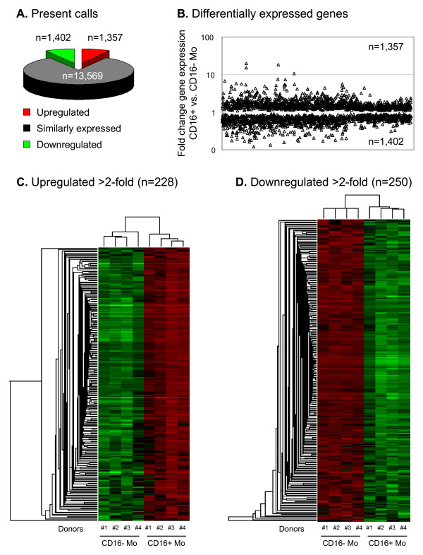
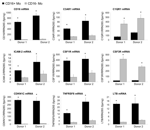
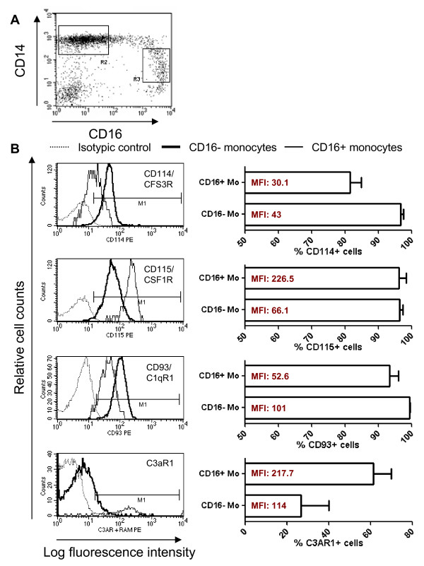
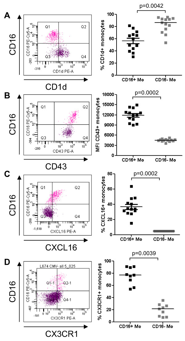
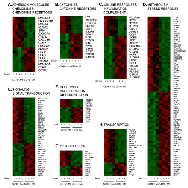
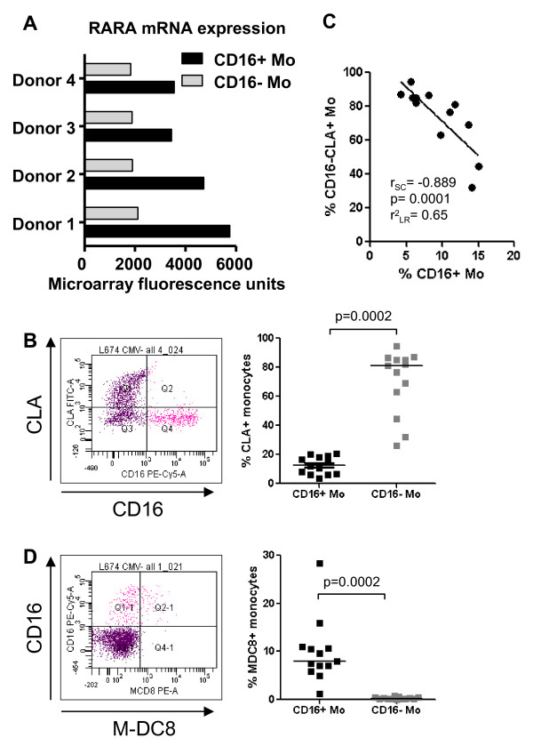
Results: To gain insight into the developmental relationship and functions of CD16+ and CD16- Mo, we examined transcriptional profiles of these Mo subsets in peripheral blood from healthy individuals. Of 16,328 expressed genes, 2,759 genes were differentially expressed and 228 and 250 were >2-fold upregulated and downregulated, respectively, in CD16+ compared to CD16- Mo. CD16+ Mo were distinguished by upregulation of transcripts for dendritic cell (DC) (SIGLEC10, CD43, RARA) and macrophage (MPhi) (CSF1R/CD115, MafB, CD97, C3aR) markers together with transcripts relevant for DC-T cell interaction (CXCL16, ICAM-2, LFA-1), cell activation (LTB, TNFRSF8, LST1, IFITM1-3, HMOX1, SOD-1, WARS, MGLL), and negative regulation of the cell cycle (CDKN1C, MTSS1), whereas CD16- Mo were distinguished by upregulation of transcripts for myeloid (CD14, MNDA, TREM1, CD1d, C1qR/CD93) and granulocyte markers (FPR1, GCSFR/CD114, S100A8-9/12). Differential expression of CSF1R, CSF3R, C1QR1, C3AR1, CD1d, CD43, CXCL16, and CX3CR1 was confirmed by flow cytometry. Furthermore, increased expression of RARA and KLF2 transcripts in CD16+ Mo coincided with absence of cell surface cutaneous lymphocyte associated antigen (CLA) expression, indicating potential imprinting for non-skin homing.

Conclusion: These results suggest that CD16+ and CD16- Mo originate from a common myeloid precursor, with CD16+ Mo having a more MPhi - and DC-like transcription program suggesting a more advanced stage of differentiation. Distinct transcriptional programs, together with their recruitment into tissues via different mechanisms, also suggest that CD16+ and CD16- Mo give rise to functionally distinct DC and MPhi in vivo.


Figures

Figure 1
Figure 1
Genome wide transcriptome analysis identifies new markers for CD16+ and CD16- monocyte (Mo) subsets. (A) Total RNA from matched CD16+ and CD16- Mo samples isolated from 4 different healthy donors were reverse transcribed and hybridized on GeneChip® Human Genome U133 Plus 2.0 Arrays (Affymetrix). Statistical analyses using one way ANOVA was performed to identify differentially expressed genes (p < 0.05). Graph depicts the number of probe sets shared or differentially expressed between CD16+ and CD16- Mo. (B) Graph depicts the fold change expression of probe sets differentially expressed in CD16+ versus CD16- Mo. (C-D) Hierarchical clustering analysis based on c-fuzzy means separated the 8 samples in 2 groups that perfectly matched CD16+ and CD16- Mo; heat maps were generated using differentially expressed genes (>2-fold). Red and green signify increased and decreased gene expression, respectively.
Figure 2
Figure 2
Real-time RT-PCR validation of microarray results. The expression of CD16, C3AR1, C1QR1, ICAM-2, CSFR1, CSF3R, CDKN1C, TNFRSF8, and LTB mRNA was quantified by SYBR Green real time RT-PCR in CD16+ and CD16- Mo. The concentration of each gene was normalized to the 28S rRNA internal control and expressed as fgs RNA of a target gene per 1 ng rRNA28S. Depicted are results (mean ± SD of triplicate wells; *, p < 0.05, unpaired t-test, CD16+ versus CD16- Mo) obtained with matched cells from 2 different healthy donors.
Figure 3
Figure 3
Differential expression of CD114/CSF3R, CD115/CSF1R, CD93/C1qR1 and C3aR1 on CD16+ and CD16- monocytes. Freshly isolated PBMC were stained with FITC CD14, PE-Cy5 CD16, and PE CD114, PE CD115, and PE CD93 Abs. The expression of CD3aR1 was detected after staining with unconjugated mouse C3AR1 Ab and PE rat anti-mouse Ab (RAM). CD14highCD16neg (R2) and CD14lowCD16+ (R3) Mo (A) were analyzed for expression of CD114, CD115, CD93 and C3aR1 (B). Shown is an overlay histogram from one representative donor of 4 donors examined (B, left panels) and graphs showing mean ± SEM for % or MFI of CD114, CD115, CD93 and C3aR1 expression on each Mo subset (B, right panels). (*, Paired t-test p-value < 0.05, CD16+ versus CD16- Mo; n = 4).
Figure 4
Figure 4
Differential expression of CD1d, CD43, CXCL16, and CX3CR1 on CD16+ and CD16- monocytes. Freshly isolated PBMC were stained with Pacific Blue CD3, Alexa700 CD4, FITC CD14, PE-Cy5 CD16, and PE CD1d, PE CD43, PE CXCL16 or PE CX3CR1 Abs. Gated CD3-CD4lowCD14highCD16- (CD16- Mo) and CD3-CD4lowCD14lowCD16+ (CD16+ Mo) cells were analyzed for expression of (A) CD1d, (B) CD43, (C) CXCL16, and (D) CX3CR1. Shown are representative dot plots (left panels) and results for 9–13 different donors (right panels). Paired Wilcoxon signed rank test was used to calculated statistical significance (p < 0.05, CD16+ versus CD16- Mo).
Figure 5
Figure 5
Biological functions of genes differentially expressed in CD16+ and CD16- monocytes. Differentially expressed genes were classified based on their biological functions using Gene Ontology as indicated. Heat maps were generated using dChip software and include data from matched CD16+ and CD16- Mo from 4 different individuals. In each heat map, upregulated genes are plotted first followed by downregulated genes. Red and green signify increased and decreased gene expression, respectively.
Figure 6
Figure 6
Differential expression of RARA mRNA and PSGL-1 epitopes CLA and M-DC8 on CD16+ and CD16- monocytes. (A) Differential expression of RARA mRNA in CD16+ and CD16- Mo was extracted from microarray data set results and expressed as relative fluorescence units. (B-D) Freshly isolated PBMC were stained with Pacific Blue CD3, Alexa700 CD4, PE-Cy5 CD16, FITC CLA and PE M-DC8 Abs. Gated CD3-CD4lowCD14highCD16- (CD16- Mo) and CD3-CD4lowCD14lowCD16+ (CD16+ Mo) cells were analyzed for expression of CLA (C) and M-DC8 (D). (B and D) Shown are representative dot plots (left panels) and results for 13 different donors (right panels). Paired Wilcoxon signed rank test was used to calculated statistical significance (p < 0.05). (B) Spearman correlation (r and p values) and linear regression (r2 value) were calculated to examine the relationship between the frequency of CD16-CLA+ Mo and CD16+ Mo.

References

    1. Randolph GJ, Inaba K, Robbiani DF, Steinman RM, Muller WA. Differentiation of phagocytic monocytes into lymph node dendritic cells in vivo. Immunity. 1999;11:753–761. doi: 10.1016/S1074-7613(00)80149-1. - DOI - PubMed
    1. Serbina NV, Pamer EG. Monocyte emigration from bone marrow during bacterial infection requires signals mediated by chemokine receptor CCR2. Nat Immunol. 2006;7:311–317. doi: 10.1038/ni1309. - DOI - PubMed
    1. Fogg DK, Sibon C, Miled C, Jung S, Aucouturier P, Littman DR, Cumano A, Geissmann F. A clonogenic bone marrow progenitor specific for macrophages and dendritic cells. Science. 2006;311:83–87. doi: 10.1126/science.1117729. - DOI - PubMed
    1. Geissmann F. The origin of dendritic cells. Nat Immunol. 2007;8:558–560. doi: 10.1038/ni0607-558. - DOI - PubMed
    1. Varol C, Landsman L, Fogg DK, Greenshtein L, Gildor B, Margalit R, Kalchenko V, Geissmann F, Jung S. Monocytes give rise to mucosal, but not splenic, conventional dendritic cells. J Exp Med. 2007;204:171–180. doi: 10.1084/jem.20061011. - DOI - PMC - PubMed

Publication types