Review
. 2009 Aug 31;168(1-2):59-68.
doi: 10.1016/j.resp.2009.02.001. Epub 2009 Feb 13.

Retrotrapezoid nucleus, respiratory chemosensitivity and breathing automaticity

Affiliations
Review

Retrotrapezoid nucleus, respiratory chemosensitivity and breathing automaticity

Patrice G Guyenet et al. Respir Physiol Neurobiol. .

Abstract

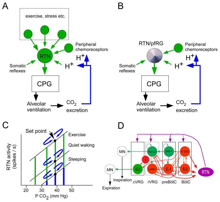
Breathing automaticity and CO(2) regulation are inseparable neural processes. The retrotrapezoid nucleus (RTN), a group of glutamatergic neurons that express the transcription factor Phox2b, may be a crucial nodal point through which breathing automaticity is regulated to maintain CO(2) constant. This review updates the analysis presented in prior publications. Additional evidence that RTN neurons have central respiratory chemoreceptor properties is presented, but this is only one of many factors that determine their activity. The RTN is also regulated by powerful inputs from the carotid bodies and, at least in the adult, by many other synaptic inputs. We also analyze how RTN neurons may control the activity of the downstream central respiratory pattern generator. Specifically, we review the evidence which suggests that RTN neurons (a) innervate the entire ventral respiratory column and (b) control both inspiration and expiration. Finally, we argue that the RTN neurons are the adult form of the parafacial respiratory group in neonate rats.


Figures

Figure 1
Figure 1. Role of RTN in breathing: a working hypothesis
A: In the adult, the RTN no longer possesses intrinsic pacemaker properties (Mulkey et al., 2004). We postulate that the discharge of RTN neurons is regulated by their “chemical drive” (intrinsic pH-sensitivity and inputs from peripheral chemoreceptors) and by synaptic inputs that develop along with various behaviors that require large changes in respiration (emotions, stress and particularly physical exercise). B: In the neonate the RTN, a subset of neurons previously identified as the pfRG, is a cluster of pH-sensitive excitatory neurons endowed with intrinsic bursting activity (Onimaru et al., 2008). We hypothesize that the intrinsic bursting properties of these neurons provide a background activity that enables these cells to be up and down regulated by the local pH and, presumably, also by inputs from peripheral chemoreceptors. C: graphic representation of how RTN neurons might regulate arterial PCO2 in the adult. The chemical drive of the RTN neurons is represented in blue and the other drives (synaptic drives from within the brain) in green. Any small deviation of PCO2 from the set-point translates into an increase or decrease in the chemical drive of the RTN cells; the effect is to increase or decrease ventilation and bring PCO2 back to the set-point (mechanism represented by the curved blue arrows). When metabolic requirements are low, the chemical drive may be the predominant factor that maintains the activity of the RTN neurons. D: theoretical concept of how RTN might differentially control the respiratory rhythm, the inspiratory motor outflow and the activity of expiratory muscles. The core components of the rhythm and pattern generating circuit are redrawn after Rybak et al. ( 2007). Excitatory neurons that presumably function via fast ionotropic transmission are in green. The pre-I glutamatergic neurons are the primary oscillator of the preBötC (Feldman and Del Negro, 2006). Inhibitory neurons that use ionotropic GABAergic or glycinergic transmission are in red (Rybak et al., 2007). RTN neurons are depicted in magenta to convey the notion that their post-synaptic effect may be mediated by a slow signaling pathway (“tonic drive”, Rybak et al. ( 2007)). Expiratory pre-motor neurons (E2, i.e. E-AUG) are located in the cVRG. These neurons derive their expiratory-augmenting membrane trajectory from inhibitory volleys (GABA or glycine) during the I and the post-I (E-DEC) phases of the respiratory cycle (Iscoe, 1998). To account for evidence that the RTN region regulates expiration (Janczewski and Feldman, 2006b), we hypothesize that a subset of RTN neurons provides a form of tonic excitatory drive to the expiratory pre-motor neurons of the cVRG (E2, green). RTN neurons collectively innervate the cVRG, rVRG, preBötC and BötC regions but the exact neuronal targets within these regions are unknown and it is also unknown whether the cVRG, rVRG, preBötC and BötC are differentially innervated by subsets of RTN neurons. Some of the ramp-I premotor neurons located in the dorsal respiratory group (not represented) may also be targeted by RTN neurons which innervate this region of the NTS. Finally, RTN neurons have a respiratory modulation with an intensity roughly proportional to that of the central respiratory drive (Guyenet et al., 2005a). This modulation could be a form of feedback that originates from inhibitory interneurons with early-I, E-DEC and E-2 activity (Guyenet et al., 2005a). The location of these inhibitory neurons in the ventral respiratory group where indicated is only a guess.

Similar articles

Cited by

References

    1. Alheid GF, McCrimmon DR. The chemical neuroanatomy of breathing. Respir Physiol Neurobiol 2008 - PMC - PubMed
    1. Amiel J, Laudier B, Attie-Bitach T, Trang H, de PL, Gener B, Trochet D, Etchevers H, Ray P, Simonneau M, Vekemans M, Munnich A, Gaultier C, Lyonnet S. Polyalanine expansion and frameshift mutations of the paired-like homeobox gene PHOX2B in congenital central hypoventilation syndrome. Nat Genet. 2003;33:459–461. - PubMed
    1. Arata A, Onimaru H, Homma I. Possible synaptic connections of expiratory neurons in the medulla of newborn rat in vitro. NeuroReport. 1998;9:743–746. - PubMed
    1. Ballanyi K, Onimaru H, Homma I. Respiratory network function in the isolated brainstem-spinal cord of newborn rats. Prog Neurobiol. 1999;59:583–634. - PubMed
    1. Bayliss DA, Talley EM, Sirois JE, Lei QB. TASK-1 is a highly modulated pH-sensitive ‘leak’ K+ channel expressed in brainstem respiratory neurons. Resp Physiol. 2001;129:159–174. - PubMed

Publication types