. 2009 Oct;30(10):869-75.
doi: 10.1016/j.placenta.2009.08.003. Epub 2009 Aug 26.

Differential regulation of human PlGF gene expression in trophoblast and nontrophoblast cells by oxygen tension

Affiliations

Differential regulation of human PlGF gene expression in trophoblast and nontrophoblast cells by oxygen tension

R M Gobble et al. Placenta. 2009 Oct.

Abstract

Objective: To determine the mechanism for differential effects of low oxygen tension on human PlGF gene transcription in trophoblast and nontrophoblast cells.

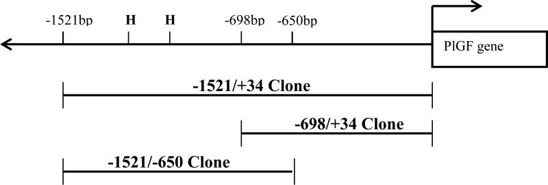
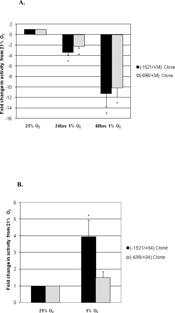
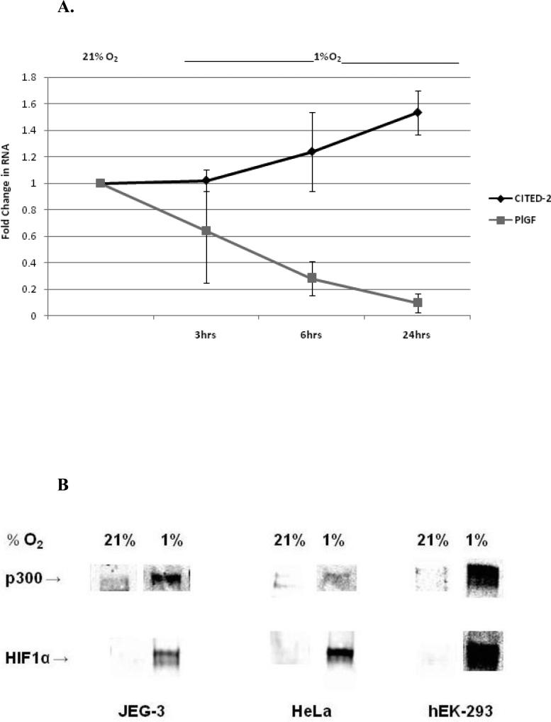
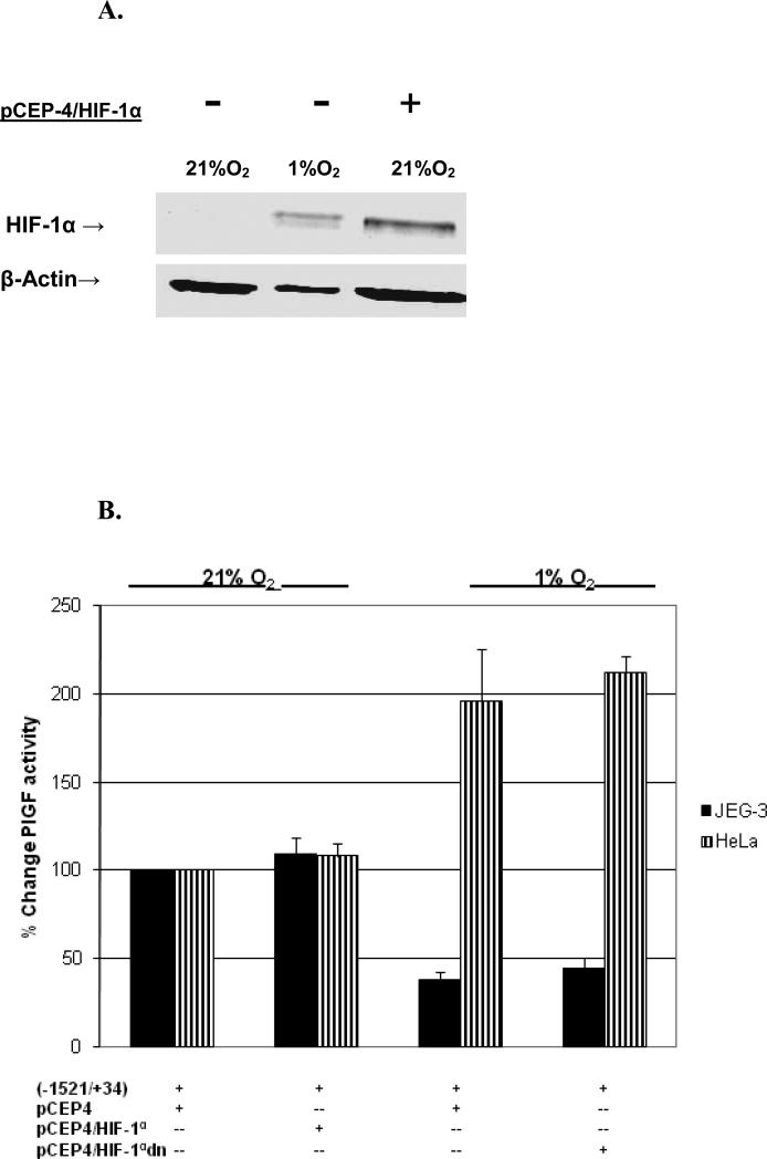
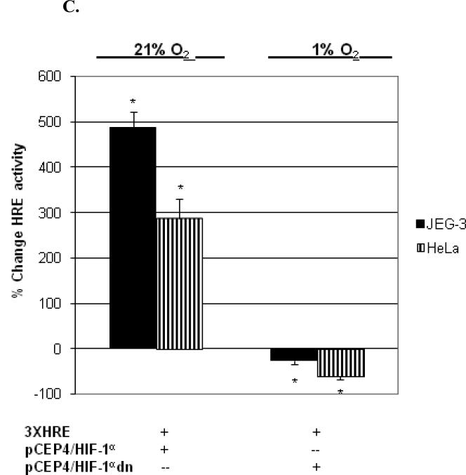
Study design: Human PlGF reporter clones and real-time RT-PCR were used to compare the effects of hypoxia on gene transcription in human trophoblast and nontrophoblast cell lines. Overexpression of HIF-1alpha, inhibition of HIF-1 function and biochemical assessments of HIF-1 co-factor interactions were used to characterize hypoxia response mechanisms regulating PlGF transcription.

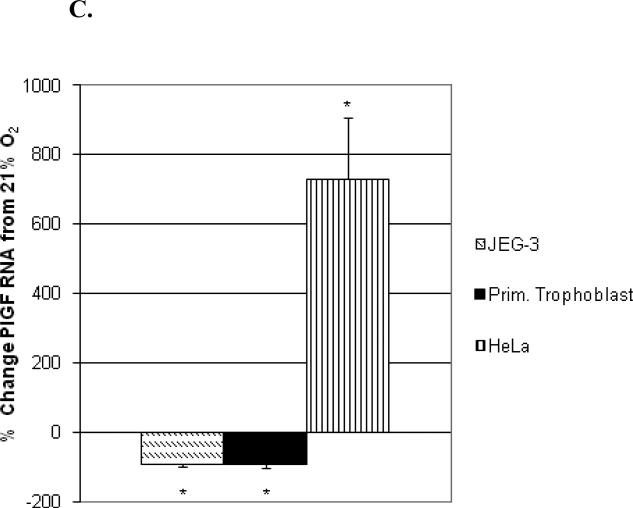
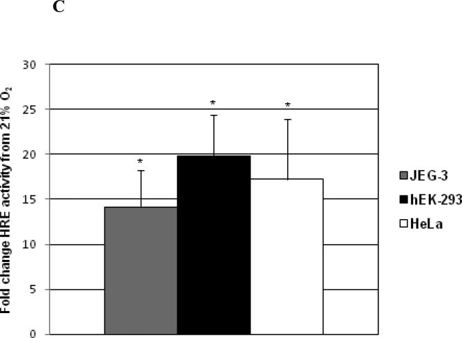
Results: PlGF transcription is specifically inhibited by low oxygen tension in trophoblast but is induced in some nontrophoblast cells. Overexpression of HIF-1alpha in normoxic cells or inhibition of HIF-1 function in hypoxic cells did not significantly alter transcription patterns of the PlGF gene in either cell type.

Conclusions: These results suggest that transcriptional repression of PlGF gene expression occurs in human trophoblast exposed to low oxygen tension but that PlGF transcription is stimulated in certain hypoxic nontrophoblast cells. However, regulation of PlGF transcription is not mediated by functional HIF-1 activity in either cell type.


Figures

Figure 1
Figure 1. Schematic of PlGF Promoter Clones
Specific regions of the 5’ flanking region of the human PlGF gene were PCR amplified, ligated into pBlueTOPO and verified by bidirectional sequencing and alignment with the published human genome sequence for each region as detailed previously [27]. H = consensus HIF-1 response elements (HRE).
Figure 2
Figure 2. Differential effects of oxygen tension on PlGF transcription and mRNA expression in trophoblast versus nontrophoblast
(A) Hypoxia significantly decreases transcription of PlGF promoter clones in trophoblast. JEG-3 cells were co-transfected with RSV-luc and PlGF promoter clones (-1521/+34) or (-698/+34). Fold decrease of β-gal activity under 1%O2 conditions from β-gal activity in 21%O2 is plotted (± SEM). All decreases in transcriptional activity were statistically significant (* = p < 0.01) from 21% O2 levels (n ≥ 5 independent experiments). (B) Hypoxia increases transcription of (-1521/+34) PlGF clone in HeLa cells. Cells were co-transfected as in panel A and fold increase in β-gal activity under 1% O2 conditions compared to 21% O2 normoxic values plotted , * = p < 0.02, n = 9 independent experiments. (C) Cell type specific effects of hypoxia on PlGF mRNA expression. Primary trophoblast, JEG-3, and HeLa cells were cultured under 21% O2 or 1% O2 for 24hrs. PlGF mRNA expression was determined by real-time RT-PCR. Data is plotted as mean percent change from normoxic culture conditions (21% O2 = 100%), * = p ≤ 0.05.
Figure 3
Figure 3. Hypoxia induces similar HIF-1α expression and nuclear translocation between cell types
(A). Cells were subjected to culture conditions at 21% O2 or 1% O2 for 24 hrs. 40μg of nuclear protein lysates were prepared and subjected to Western blot analysis for HIF-1α. Membranes were stripped and re-probed for β-actin to normalize for protein loading. (n = 3).
Figure 4
Figure 4. Hypoxia induces CITED-2 expression and similar HIF-1α translocation, p300 recruitment, and functional HIF-1 activity
(A). Hypoxia induces CITED-2 mRNA expression and decreases PlGF expression in trophoblast. JEG-3 cells were subjected to DFO (3hr) or 1% O2 for 6 and 24hrs. Relative change in mRNA expression for each time point was determined by real-time RT-PCR and normalized to levels at 21% O2 (set = 1). (B) Nuclear HIF-1α in hypoxic cells binds to consensus HRE and recruits p300. Nuclear protein lysates were mixed with Avidin-D-HRE beads and proteins bound to the HRE-oligo were subjected to Western blot analysis for p300 and HIF-1α. Representative blots from 5-7 independent assays are shown. (C) Hypoxia induces similar HIF-1 functional activity in trophoblast and nontrophoblast cell lines. Cells were co-transfected with pSV-β-gal and pGL2-TK-HRE, and cultured in 21% O2 or 1% O2 conditions. Fold increase of luciferase activity under hypoxic conditions above activity in 21% O2 is plotted, n = 6 independent experiments/cell line, p<0.05 from normoxic values for each cell line.
Figure 5
Figure 5. Transcriptional activity of PlGF promoter (-1521/+34) under normoxic or hypoxic cell culture conditions is independent of HIF-1α
(A.) JEG-3 cells were transfected with pCEP-4/HIF-1α, subjected to 21% O2 or 1% O2 for 24hrs, and nuclear proteins immunoblotted for HIF-1α and β-actin. (B and C.) JEG-3 and HeLa cells were co-transfected with PlGF clone (-1521/+34) (panel B) or pGL2-TK-HRE (panel C) reporter clone and either pCEP-4, pCEP-4/HIF-1α or pCEP-4/HIF-1αdn. Cells were lysed after 24 hrs in 21% O2 or 1% O2 conditions and normalized percent change in reporter activities from control transfections plotted with *= < 0.05 (n=5).

Similar articles

Cited by

References

    1. Ziche M, Maglione D, Ribatti D, Morbidelli L, Lago CT, Battisti M, Paoletti I, Barra A, Tucci M, Parise G, Vincenti V, Granger HJ, Viglietto G, Persico MG. Placenta growth factor-1 is chemotactic, mitogenic, and angiogenic. Lab Invest. 1997;76:517–31. - PubMed
    1. Maglione D, Guerriero V, Viglietto G, Delli-Bovi P, Persico MG. Isolation of a human placenta cDNA coding for a protein related to the vascular permeability factor. Proc Natl Acad Sci U S A. 1991;88:9267–71. - PMC - PubMed
    1. Khaliq A, Li XF, Shams M, Sisi P, Acevedo CA, Whittle MJ, Weich H, Ahmed A. Localisation of placenta growth factor (PIGF) in human term placenta. Growth Factors. 1996;13:243–50. - PubMed
    1. Clark DE, Smith SK, Licence D, Evans AL, Charnock-Jones DS. Comparison of expression patterns for placenta growth factor, vascular endothelial growth factor (VEGF), VEGF-B and VEGF-C in the human placenta throughout gestation. J Endocrinol. 1998;159:459–67. - PubMed
    1. Hanna J, Goldman-Wohl D, Hamani Y, Avraham I, Greenfield C, Natanson-Yaron S, Prus D, Cohen-Daniel L, Arnon TI, Manaster I, Gazit R, Yutkin V, Benharroch D, Porgador A, Keshet E, Yagel S, Mandelboim O. Decidual NK cells regulate key developmental processes at the human fetal-maternal interface. Nat Med. 2006;12:1065–1074. - PubMed

Publication types

MeSH terms