. 2009 Sep;32(9):496-505.
doi: 10.1016/j.tins.2009.05.010. Epub 2009 Aug 25.

From selective vulnerability to connectivity: insights from newborn brain imaging

Affiliations

From selective vulnerability to connectivity: insights from newborn brain imaging

Steven P Miller et al. Trends Neurosci. 2009 Sep.

Abstract

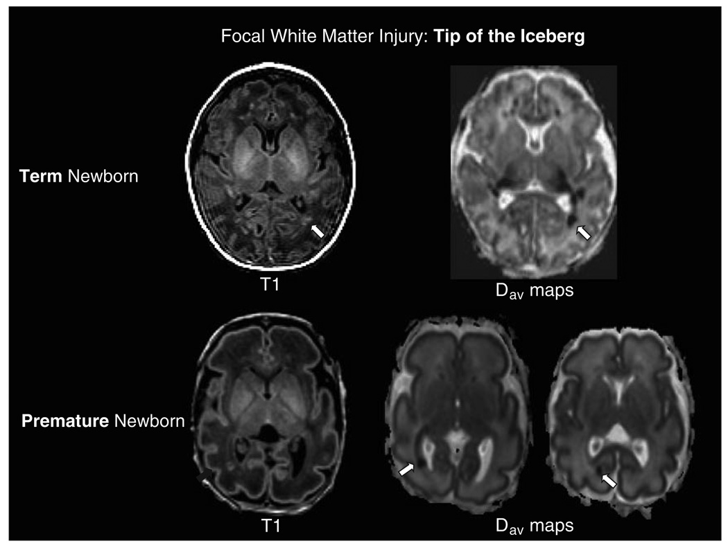
The ability to image the newborn brain during development has provided new information regarding the effects of injury on brain development at different vulnerable time periods. Studies in animal models of brain injury correlate beautifully with what is now observed in the human newborn. We now know that injury at term primarily results in grey matter injury while injury in the premature brain predominantly results in a pattern of white matter injury, though recent evidence suggests a blurring of this distinction . These injuries affect how the brain matures subsequently and again, imaging has led to new insights that allow us to match function and structure. This review will focus on these patterns of injury that are so crucially determined by age at insult. In addition, this review will highlight how the brain responds to these insults with changes in connectivity that have profound functional consequences.


Figures

Figure 1
Figure 1
A. Brain development from early in premature life to term-equivalent age. Axial T2 weighted and color coded fractional anisotropy maps in a premature newborn delivered at 28 weeks gestation and scanned at 30 weeks postmenstrual age and again at 39 weeks. On the T2 weighted images note the dramatic increase in gyration of the cerebral cortex and early myelination in the posterior limb of the internal capsule. The color coded fractional anisotropy maps represent the “directionality” of water diffusion, with brighter areas indicating more directionality (higher FA). Water diffusion in the right-left plane is coloured red, superior-inferior in blue, and anterior –posterior in green. Note the loss of FA in the cerebral cortex from preterm to term-equivalent age, consistent with the normal loss of cortical radial organization. White matter tracts such as the corticospinal tracts (blue region in the posterior limb of the internal capsule) and optic radiations (green tract from the thalamus to calcarine cortex) are more clearly delineated with higher FA at term-equivalent age. B. Predominant Patterns of Brain Injury in Preterm and Term Newborns. In the preterm newborn delivered at 28 weeks gestation and scanned at 30 weeks postmenstrual age, the axial image from the spoiled gradient echo volumetric scan demonstrates several foci of abnormal T1 hyperintensity in the periventricular white matter consistent with focal non-cystic white matter injury (blue arrows; note lesions are evident bilaterally). In the term newborn with encephalopathy following acute profound asphyxia, the axial image from the T1-weighted sequence on the tenth day of life demonstrates abnormal T1 hyperintensity in the basal nuclei and perirolandic cortex in the characteristic basal nuclei predominant pattern of injury (yellow arrows; note abnormalities are evident bilaterally).
Figure 2
Figure 2. Patterns of Brain Injury
Deep Nuclei and Watershed Patterns of Brain Injury (A–C): MRI scan demonstrating the basal nuclei predominant pattern of injury (from Figure 1). Neuropathology from the first day of life in a term newborn following acute profound asphyxia. On gross pathology (A) injury is evident as brown discoloration in the thalami, basal ganglia, hippocampi, and cerebral cortex in a vascular watershed distribution (arrow). On hematoxylin and eosin stained microscopic sections, diffuse neuronal injury in the thalamus (B) is evident with eosinophilic (dead) neurons (arrowhead), and in the cerebellar cortex (C) with eosinophilic Purkinje cells (arrowhead). White Matter Injury (D–F): MRI scan demonstrating white matter injury (from Figure 1). Neuropathology from the second month of life demonstrating the cystic and diffuse components of periventricular leukomalacia. On gross pathology (D) cystic degeneration is present in the periventricular white matter injury (arrow). On hematoxylin and eosin stained microscopy sections, the cyst (E) (arrowhead) contains macrophages and is surrounded by marked astrogliosis and calcifications. In a section of white matter remote from the cyst (F), there is a marked paucity of myelin and astrogliosis. Images courtesy of Dr. Glenda Hendson (University of British Columbia).
Figure 3
Figure 3
Focal White Matter Injury- Tip of the Iceberg. Term Newborn: Axial images from the T1-weighted sequence and the average diffusivity maps (Dav) in a term newborn with encephalopathy and white matter injury. In addition to the discreet focus of white matter injury in the optic radiations on the T1-weighted image (arrow), note the more extensive abnormality of restricted diffusion (hypointensity on the average diffusivity map, arrow) extending from the focal white matter lesion along the optic radiations to the thalamus. Similar findings are evident with a smaller lesion contralaterally. Premature Newborn: Axial images from a premature newborn at 31 weeks postmenstrual age demonstrate a focus of white matter injury in the optic radiations on the T1-weighted spoiled gradient echo volumetric sequence. The axial average diffusivity images demonstrate the focus of white matter injury as an area of restricted diffusion (hypointensity, arrow) accompanied by extension of the restricted diffusion along the optic radiations from the thalamus to the calcarine cortex (arrow). These images highlight the effects of focal white matter lesions on “connected” white matter pathways such as the optic radiations-from the thalamus to the calcarine cortex.
Figure 4
Figure 4
(A) Neural Network Degeneration: Fiber tracking, overlayed on direction encoded fractional anisotropy maps, reveals significant connection with axons entering the septum in a control C57BL6 mouse images on postnatal day 42 (i and iii). Following neonatal hypoxia ischemia at postnatal day seven in a C57BL6 mouse (ii and iv), almost no ipsilateral fimbria connections remain through the ipsilateral column of fornix to the septum (yellow tracks) while robust contralateral axonal connections remain (red tracks). (B)Imaging Connectivity in Neonatal Stroke: Following stroke in the neonatal period (i), a minimal decrease in fractional anisotropy (FA) in the ipsilateral posterior limb of the internal capsule (PLIC) and cerebral peduncle (CP) is seen at birth, with complete loss of FA at 3 months (white arrows) (FA: fractional anisotropy map; DEC: Direction encode color map). Note the loss of high signal on the FA map in the position of the PLIC, also evident as a loss of blue signal (superior-inferior tracks) on the DEC map. When the 3 month scan is mapped against templated uninjured controls (ii), the deficit in the ipsilateral PLIC and CP is evident (red signal at arrow). Severe pruning of ipsilateral fibers (yellow) tracking through the PLIC and cerebral peduncle at 3 months of age (iii) portends diagnosis of severe hemiplegia at 2 years of age. Fiber tracking in another patient shows mild differences at 3 months; this patient developed a milder gross motor deficit (iv). Images courtesy of Dr. Frances Northington.

References

    1. Marlow N, Rose AS, Rands CE, et al. Neuropsychological and educational problems at school age associated with neonatal encephalopathy. Arch Dis Child Fetal Neonatal Ed. 2005;90:F380–F387. - PMC - PubMed
    1. Gonzalez FF, Miller SP. Does perinatal asphyxia impair cognitive function without cerebral palsy? Arch Dis Child Fetal Neonatal Ed. 2006;91:F454–F459. - PMC - PubMed
    1. van Handel M, Swaab H, de Vries LS, et al. Long-term cognitive and behavioral consequences of neonatal encephalopathy following perinatal asphyxia: a review. Eur J Pediatr. 2007;166:645–654. - PMC - PubMed
    1. Ment LR, Bada HS, Barnes P, et al. Practice parameter: neuroimaging of the neonate: report of the Quality Standards Subcommittee of the American Academy of Neurology and the Practice Committee of the Child Neurology Society. Neurology. 2002;58:1726–1738. - PubMed
    1. Schouman-Claeys E, Henry-Feugeas MC, Roset F, et al. Periventricular leukomalacia: correlation between MR imaging and autopsy findings during the first 2 months of life. Radiology. 1993;189:59–64. - PubMed

Publication types