. 2009 Oct 16;284(42):28599-606.
doi: 10.1074/jbc.M109.047282. Epub 2009 Aug 27.

Regulation of immature dendritic cell migration by RhoA guanine nucleotide exchange factor Arhgef5

Affiliations

Regulation of immature dendritic cell migration by RhoA guanine nucleotide exchange factor Arhgef5

Zhenglong Wang et al. J Biol Chem. .

Abstract

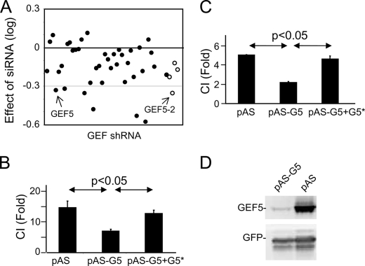
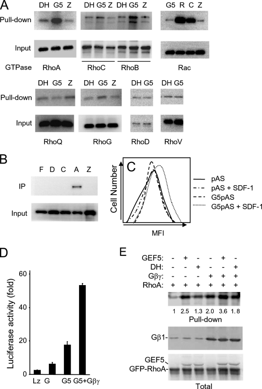
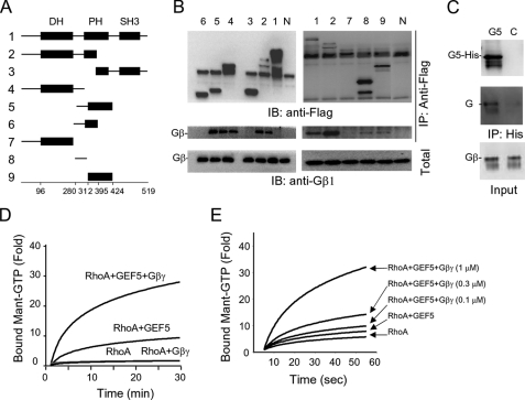
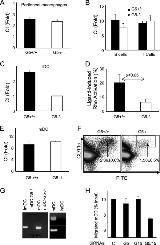
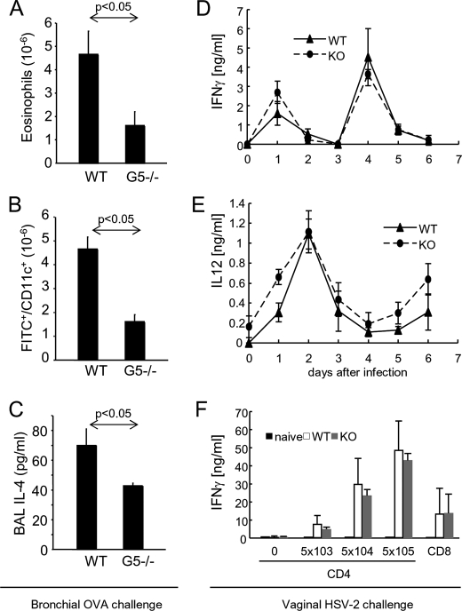
There are a large number of Rho guanine nucleotide exchange factors, most of which have no known functions. Here, we carried out a short hairpin RNA-based functional screen of Rho-GEFs for their roles in leukocyte chemotaxis and identified Arhgef5 as an important factor in chemotaxis of a macrophage phage-like RAW264.7 cell line. Arhgef5 can strongly activate RhoA and RhoB and weakly RhoC and RhoG, but not Rac1, RhoQ, RhoD, or RhoV, in transfected human embryonic kidney 293 cells. In addition, Gbetagamma interacts with Arhgef5 and can stimulate Arhgef5-mediated activation of RhoA in an in vitro assay. In vivo roles of Arhgef5 were investigated using an Arhgef-5-null mouse line. Arhgef5 deficiency did not affect chemotaxis of mouse macrophages, T and B lymphocytes, and bone marrow-derived mature dendritic cells (DC), but it abrogated MIP1alpha-induced chemotaxis of immature DCs and impaired migration of DCs from the skin to lymph node. In addition, Arhgef5 deficiency attenuated allergic airway inflammation. Therefore, this study provides new insights into signaling mechanisms for DC migration regulation.


Figures

FIGURE 1.
FIGURE 1.
Effects of GEF shRNAs on chemotaxis. A, effects of the GEF shRNAs on RAW264.7 cell chemotaxis in response to C5a. The chemotactic index of cells expressing pAS is taken as 1, and the relative chemotactic activity of cells expressing each GEF shRNA was calculated. Data shown are log of the relative chemotactic activities. The raw data are shown in supplemental Table S1. The open circles denote the second shRNAs used for validation of the initial hits. B and C, rescuing the effect of Arhgef5 shRNA by expressing an Arhgef5 mutant containing a silent mutation. Raw 264.7 cells (B) and J774 cells (C) were cotransfected with pAS, Arhgef5 shRNA (pAS-G5), and/or silently mutated Arhgef5 (G5*) for 48 h. Chemotactic assays were carried out using the transwell plate in the presence of C5a for RAW264.7 cells and SDF-1 for J774 cells. CI stands for chemotactic index. D, Arhgef5 shRNA validation. HEK293T cells were cotransfected with the plasmids expressing Arhgef5 and GFP and pAS or pAS-G5. Western analyses were carried out 2 days after transfection.
FIGURE 2.
FIGURE 2.
Regulation of Rho small GTPases by Arhgef5. A, pulldown assays. HEK293T cells were cotransfected with one of the small GTPases and LacZ (Z), Arhgef5 (G5), or the loss-of-function mutant of Arhgef5 (DH). The RBD pulldown assays were carried out for cells expressing RhoA, RhoB, and RhoC, whereas the PBD pulldown assay was done for Rac, RhoQ, RhoD, and RhoV. The activity of RhoG was determined by a pulldown using GST-Elmo. Both precipitated and total GTPases were detected by Western analysis using an antibody specific for the HA tag carried by these small GTPases. B, interaction of dominant negative GTPases with Arhgef5. HA-tagged RhoA-N19 (A), RhoC-N19 (C), RhoD-N31 (D), RhoF-N33 (F), and LacZ were cotransfected with FLAG-tagged Arhgef5 in HEK293T cells. Immunoprecipitation (IP) was carried out with an anti-HA antibody and detected with an anti-FLAG antibody. C, knocking down Arhgef5 reduced SDF-1-induced RBD binding. J774 cells were transfected with pAS or Arhgef5 shRNA for 48 h and stimulated with or without 30 ng/ml SDF-1 for 15 s. Cells were fixed, permeabilized, stained with GST-RBD and Alexa 633-labeled secondary antibody, and analyzed by a flow cytometer. MFI of Alexa 633 in cells gated for GFP is shown. Three experiments were performed. A representative one is shown. D, SRE.L-luciferase assay. HEK293T cells were co-transfected with the SRE-luciferase reporter gene, GFP, LacZ, Gβγ, and/or Arhgef5. Cells were lysed and the GFP levels and luciferase activity were determined. The luciferase activity was normalized against the GFP level. E, activation of RhoA by Gβγ and Ahrgef5 in a cotransfection assay. HEF293T cells were transfected as indicated. Twenty-four hours later, RBD pulldown assays were carried out. The numbers under the top panel are relative band intensity quantified by densitometry and normalized against total RhoA levels.
FIGURE 3.
FIGURE 3.
Gβγ interacts with and directly activates Arhgef5. A, schematic representation of Arhgef5 and its mutants. The diagrams are not drawn in scale. The numbers at the side correspond to those in B. B, interaction of Gβγ with Arhgef5 and its deletion mutants. FLAG-tagged Arhgef5 and its mutant were cotransfected with Gβ1γ2 in HEK293T cells. Immunoprecipitation (IP) was carried out using the anti-FLAG antibody and detected using an anti-Gβ1 antibody. N, no Arhgef5 transfected. C, direct interaction between Arhgef5 and Gβ1γ2. His-tagged Arhgef5 protein purified from a bacterial expression system was incubated with Gβγ from a baculoviral expression system. Pulldown was carried out by an anti-His antibody and detected by an anti-Gβ1 antibody. D, direct regulation of Arhgef5 by Gβ1γ2. Purified recombinant proteins of Gβ1γ2 (1 μm), Arhgef5 (0.5 μm), and/or RhoA (0.12 μm) were incubated as indicated in the figure in the presence of Mant-GTP. E, dose-dependent activation of RhoA by Gβγ. Different doses of Gβ1γ2 were incubated with RhoA (0.12 μm) and Arhgef5 (0.5 μm). IB, immunoblot.
FIGURE 4.
FIGURE 4.
Arhgef5 has an important role in immature DC migration. A–C, transwell migration assays. Chemotactic activity of peritoneal macrophages (A), spleen T and B lymphocytes (B), and bone marrow-derived immature DCs (C) from wild type (G5+/+) or Arhgef5-deficient (G5−/−) mice was determined using the transwell assay. C5a (10 ng/ml, A), SDF-1 (100 ng/ml, B), and MIF1α (300 ng/ml, C) were used. D, RhoA activity in immature DCs. MIP-1α-induced Rho activation was determined using an ELISA kit that determines the levels of active RhoA. p = 0.012. E, transwell migration assay of bone marrow-derived mature DCs in response to CCL19 (60 ng/ml). F, in vivo migration of DCs. Mice were painted with FITC and cells from inguinal lymph nodes were isolated and stained with CD11c. The percentage of DCs migrated from the skins (FITC/CD11c double positive) were determined by flow cytometry. n = 14, p < 0.05. G, expression of Arhgef5 and its close homologs Arhgef15 and Ephexin-1 detected by RT-PCR in immature DCs (imDC) and mature DCs (mDC). H, knocking down of both Arhgef5 and -15 reduces migration of bone marrow-derived mature DCs in response to CCL-19. Bone marrow-derived mature DCs were transfected with no oligo (C) or synthetic siRNA oligos targeting Arhgef5 (G5), Arhgef15 (G15), or both (G5/15). p < 0.05, fourth bar versus others. P values for other bars are >0.05.
FIGURE 5.
FIGURE 5.
Effects of Arhgef5 deficiency on two in vivo disease models. A–C, effects of Arhgef5 deficiency on an OVA-induced allergic airway inflammation model. The numbers of eosinophils in BAL (A) and FITC+/CD11C+ cells in the bronchial lymph nodes (B) and the levels of IL-4 in BAL (C) were determined, n = 7. d–F, effects of Arhgef5 deficiency on a HSV-2 infection model. The levels of IFN-γ (D) and IL-12 (E) in vaginal washes were collected daily and determined. CD4+ or CD8+ T cells were isolated from iliac and inguinal lymph nodes. They were co-cultured with naïve wild type (WT) splenocytes and heat-inactivated HSV-2 of the indicated plaque-forming units (for CD4+ T cells) or 1 μg/ml gB peptide (for CD8+ T cells) for 3 days. The levels of IFN-γ in the conditioned medium were determined by ELISA (F). n = 3 (wild type) or 4 (Arhgef5−/−).

Similar articles

Cited by

References

    1. Ridley A. J. (2001) J. Cell Sci. 114, 2713–2722 - PubMed
    1. Etienne-Manneville S., Hall A. (2002) Nature 420, 629–635 - PubMed
    1. Allen W. E., Zicha D., Ridley A. J., Jones G. E. (1998) J. Cell Biol. 141, 1147–1157 - PMC - PubMed
    1. Srinivasan S., Wang F., Glavas S., Ott A., Hofmann F., Aktories K., Kalman D., Bourne H. R. (2003) J. Cell Biol. 160, 375–385 - PMC - PubMed
    1. Meili R., Firtel R. A. (2003) Cell 114, 153–156 - PubMed

Publication types