Comparative Study
. 2009 Nov;76(5):1031-43.
doi: 10.1124/mol.109.057752. Epub 2009 Aug 27.

Functional characterization of human cytochrome P450 2S1 using a synthetic gene-expressed protein in Escherichia coli

Affiliations
Comparative Study

Functional characterization of human cytochrome P450 2S1 using a synthetic gene-expressed protein in Escherichia coli

Peter H Bui et al. Mol Pharmacol. 2009 Nov.

Abstract

Human cytochrome P450 2S1 was recently identified and shown to be inducible by 2,3,7,8-tetrachlorodibenzo-p-dioxin and hypoxia. It is highly expressed in epithelial cells of tissues that are exposed to the environment and in many tumors of epithelial origin. The biological function of CYP2S1 has not yet been determined, although its possible role in carcinogen metabolism has been suggested. In this report, we investigated its ability to metabolize carcinogens. To obtain a large quantity of active enzyme for substrate screening, we overexpressed CYP2S1 in Escherichia coli (200 nM culture), using a synthetic gene approach. High-level expression allowed us to achieve purification of CYP2S1 with high specific content and purity (16 nmol/mg). Despite high-level expression, we found that CYP2S1 was not readily reduced by cytochrome P450 reductase, and thus no activity was found using NADPH. However, the oxidative activity of CYP2S1 was supported by cumene hydroperoxide or H(2)O(2), such that CYP2S1 oxidized many important environmental carcinogens, including benzo[a]pyrene, 9,10-dihydro-benzo[a]pyrene, 7,12-dimethylbenz[a]anthracene, benzo[a]pyrene-7,8-dihydrodiol, aflatoxin B1, naphthalene, and styrene, with high turnover. Most substrates tested were converted to detoxification products, except in the case of benzo[a]pyrene-7,8-dihydrodiol, which was converted into the very potent carcinogenic metabolite 7,8-dihydrodiol-trans-9,10-epoxide at a relatively efficient rate (K(m) = 12.4 +/- 2 microM, turnover = 2.3 min(-1)). This metabolite formation was also supported both in vitro and in vivo by fatty acid hydroperoxides described in the accompanying report (p. 1044). Together, these data indicate that CYP2S1 contributes to the metabolism of environmental carcinogens via an NADPH independent activity.


Figures

Fig. 1.
Fig. 1.
Comparison of the hypothetical mRNA secondary structures, ΔG values, and P450 spectral characteristics expression of native CYP2S1dh, semiCYP2S1dh, and syn2S1dh. A, structures and ΔG values were calculated at a 37°C folding temperature and 1 M Na+ using mfold (http://mfold.bioinfo.rpi.edu/cgi-bin/dna-form1.cgi). Expression of the native CYP2S1 construct (B), semiCYP2S1-dh construct (C), and syn2S1-dh construct (D) in E. coli LMG 194. All constructs were coexpressed with P450-reductase. Fe2+ CO versus Fe2+ difference spectra were measured using 1:5 dilutions of lysate supernatants. The expression levels of CYP2S1, semiCYP2S1-dh, and syn2S1-dh were estimated to be 20 to 30, 50 to 60, and 200 nM culture, respectively.
Fig. 2.
Fig. 2.
Amino acid sequence alignment of human CYP2S1 with other CYP2 family enzymes. The highlighted amino acids are thought to be important in the interaction between the cytochromes P450 and P450 reductase.
Fig. 3.
Fig. 3.
NADPH reduced Fe2+ CO versus Fe2+ difference spectra of CYP2S1 + P450 reductase (A). Light blue, CYP2S1 + CO; brown, CYP2S1 + CO + NADPH; blue, CYP2S1 + CO + NADPH + naphthalene; purple, CYP2S1 + CO + NADPH + naphthalene + dithionite. Spectra of CYP1A1 + P450 reductase (B): green, CYP1A1 + CO; blue, CYP1A1 + CO + NADPH; red, CYP1A1 + CO + NADPH + harmaline. Spectra of CYP2C9 + P450 reductase (C): black, CYP2C9 + CO; blue, CYP2C9 + CO + NADPH; light blue, CYP2C9 + CO + NADPH + diclofenac; green, CYP2C9 + CO + dithionite. Spectra of CYP3A4 + P450 reductase (D), green, CYP3A4 + CO; red, CYP3A4 + CO + NADPH; purple, CYP3A4 + CO + NADPH + diclofenac; blue, CYP3A4 + CO + NADPH + testosterone.
Fig. 4.
Fig. 4.
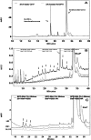
Cumene hydroperoxide and hydrogen peroxide support oxidation by CYP2S1. HPLC chromatograms of 0.15 μM CYP3A4 + reductase (A) incubated with 100 μM testosterone and 1 mM cumene hydroperoxide (dark line), or with 200 μM testosterone and a NADP/NADPH regeneration mixture (gray line) for 10 min at 37°C. The 19-min peak corresponds to a 6-β-OH-testosterone, detected at 245 nm. HPLC chromatograms (B) comparing activities of 0.15 μM CYP2S1 (solid black line), 0.15 μM CYP1A1 (gray line), and human reductase sample (HR, dotted line) with 100 μM benzo[a]pyrene in the presence of 1 mM cumene hydroperoxide for 10 min at 37°C. HPLC chromatograms (C) of 0.15 μM CYP2S1, 0.15 μM CYP1A1, and human reductase sample (HR) toward 100 μM benzo[a]pyrene in the presence of 10 mM H2O2 for 10 min at 37°C. Products, indicated by arrows, were detected at 254 nm.
Fig. 5.
Fig. 5.
Identification of CYP2S1-catalyzed products of BaP. HPLC chromatogram of 0.2 μM CYP2S1 incubated with 200 μM BaP and 1 mM CHP (solid black line). Products were identified at 254 nm using authentic standards (dotted lines), 6,12-BaP-quinone (4); 3,6-BaP-quinone (3); 1,6 benzo[a]pyrene (2), 4-hydroxy benzo[a]pyrene (4-OH-BaP) (5), and BaP-7,8-diol (1). Other products, indicated with question mark, were not identified.
Fig. 6.
Fig. 6.
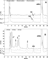
Identification of CYP2S1-catalyzed products of BaP-7,8-diol. HPLC chromatograms of 0.2 μM CYP2S1 incubated with 133 μM BaP-7,8-diol and 1 mM CHP (A), no CHP control (B), and no CYP2S1control (C). Detection was set at 345 nm. r7,t8,t9,c10-tetrol was identified using an authentic standard (D). Other unknown products, indicated by arrows, were also detected. The product marked with * has the same UV spectrum as r7,t8,t9,c10-tetrol, suggesting that it is its enantiomer.
Fig. 7.
Fig. 7.
Identification of CYP2S1-catalyzed products of 9,10-H2-BaP. HPLC chromatogram of 0.2 μM CYP2S1 incubated with 133 μM 9,10-H2-BaP and 1 mM CHP (A), without CYP2S1 (C) control, or without CHP (B) control. Detection was set at 310 nm. The 7,8-diol-9,10-H2-BaP product was identified based on the comparison of its UV-spectrum (D) to that of r7,t8,t9,c10-tetrol (E).
Fig. 8.
Fig. 8.
Identification of CYP2S1-catalyzed products of DMBA. HPLC chromatogram of 0.2 μM DMBA incubated with 500 μM DMBA and CHP (solid black). Products were identified at 295 nm using authentic standards, 12-OH-DMBA (3), 7-OH-DMBA (2), and 3,4-diol DMBA (1). Other unknown products are indicated by question marks.
Fig. 9.
Fig. 9.
Identification of CYP2S1-catalyzed products of all-trans-retinoic acid (AtR). HPLC chromatograms of 0.2 μM CYP2S1 incubated with 20 μM AtR and 1 mM CHP (A) or 1 mM H2O2 (B), and of 0.1 μM CYP2C8 + reductase incubated with 5 μM AtR and NADPH (C). Products were detected at 350 nm. 4-OH-AtR was identified based on a previous report in which CYP2C8 produced 4-OH-AtR from AtR (Nadin and Murray, 1999).
Fig. 10.
Fig. 10.
Identification of CYP2S1-catalyzed products of aflatoxin B1. HPLC chromatograms of 0.2 μM CYP2S1 incubated with 243 μM aflatoxin B1 and 3 mM H2O2 (i), of 0.2 μM CYP3A4 + reductase incubated with 243 μM aflatoxin B1 and NADPH (ii), and of CYP1A2 + reductase incubated with 243 μM aflatoxin B1 and NADPH (iii). Products were detected with UV-Vis detector at 360 nm (A) and with a fluorescence detector (B). M1 and Q1 were identified based on a previous report (Gallagher et al., 1996). Unknown products are indicated by arrows.

References

    1. Anari MR, Khan S, Jatoe SD, O'Brien PJ. (1997) Cytochrome P450 dependent xenobiotic activation by physiological hydroperoxides in intact hepatocytes. Eur J Drug Metab Pharmacokinet 22:305–310 - PubMed
    1. Baldwin RM, Shultz MA, Buckpitt AR. (2005) Bioactivation of the pulmonary toxicants naphthalene and 1-nitronaphthalene by rat CYP2F4. J Pharmacol Exp Ther 312:857–865 - PubMed
    1. Barnes HJ, Arlotto MP, Waterman MR. (1991) Expression and enzymatic activity of recombinant cytochrome P450 17 alpha-hydroxylase in Escherichia coli. Proc Natl Acad Sci U S A 88:5597–5601 - PMC - PubMed
    1. Barr DP, Martin MV, Guengerich FP, Mason RP. (1996) Reaction of cytochrome P450 with cumene hydroperoxide: ESR spin-trapping evidence for the homolytic scission of the peroxide O-O bond by ferric cytochrome P450 1A2. Chem Res Toxicol 9:318–325 - PubMed
    1. Barr DP, Mason RP. (1995) Mechanism of radical production from the reaction of cytochrome c with organic hydroperoxides. An ESR spin trapping investigation. J Biol Chem 270:12709–12716 - PubMed

Publication types

MeSH terms

LinkOut - more resources