. 2009 Oct;37(19):6400-13.
doi: 10.1093/nar/gkp705. Epub 2009 Aug 27.

The ERCC1/XPF endonuclease is required for completion of homologous recombination at DNA replication forks stalled by inter-strand cross-links

Affiliations

The ERCC1/XPF endonuclease is required for completion of homologous recombination at DNA replication forks stalled by inter-strand cross-links

Ali Z Al-Minawi et al. Nucleic Acids Res. 2009 Oct.

Abstract

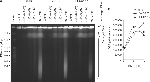
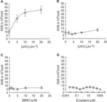
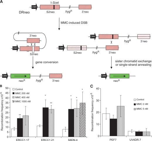
Both the ERCC1-XPF complex and the proteins involved in homoIogous recombination (HR) have critical roles in inter-strand cross-link (ICL) repair. Here, we report that mitomycin C-induced lesions inhibit replication fork elongation. Furthermore, mitomycin C-induced DNA double-strand breaks (DSBs) are the result of the collapse of ICL-stalled replication forks. These are not formed through replication run off, as we show that mitomycin C or cisplatin-induced DNA lesions are not incised by global genome nucleotide excision repair (GGR). We also suggest that ICL-lesion repair is initiated either by replication or transcription, as the GGR does not incise ICL-lesions. Furthermore, we report that RAD51 foci are induced by cisplatin or mitomycin C independently of ERCC1, but that mitomycin C-induced HR measured in a reporter construct is impaired in ERCC1-defective cells. These data suggest that ERCC1-XPF plays a role in completion of HR in ICL repair. We also find no additional sensitivity to cisplatin by siRNA co-depletion of XRCC3 and ERCC1, showing that the two proteins act on the same pathway to promote survival.


Figures

Figure 1.
Figure 1.
RAD51D cells are deficient in homologous recombination. (A) Schematic illustration of the SCneo recombination substrate. The SCneo substrate contains two non-functional copies of the neoR gene. After an I-SceI induced DSB, a functional neoR gene arises by HR via intrachromatid pairing or sister chromatid pairing. Single-strand annealing (SSA), a non-conservative HR sub-pathway, does not produce a functional neoR gene (60). (B) Recombination frequency to G418 resistance after transient transfection of the pCMV3xnlsI-SceI vector. Resistance to G418 reflects a recombination event of either intrachromatid pairing or sister chromatid pairing but not SSA. Columns depict the average and bars represent the standard deviation of at least three experiments.
Figure 2.
Figure 2.
ERCC1- and RAD51D-defective cells are hypersensitive to cross-linking agents mitomycin C and cisplatin. Survival was determined in colony outgrowth experiments in UV4DR7 (ERCC1 mutated) and ERCC1 complemented cells (ERCC1.17, ERCC1.21) or transfection with empty vector (PEF7), along with wild-type AA8 cells (AA8SN.10, AA8SN.12) or RAD51D targeted cells (51D1SC.2, 51D1SC.4) (26) as well as BRCA2-defective V-C8 cells and wild-type (V79Z) or BRCA2 complemented control (V-C8+B2) (59) following treatment with (A) mitomycin C (MMC) (B), cisplatin or (C) melphalan. The average and standard deviation of at least three experiments is depicted.
Figure 3.
Figure 3.
DNA double-strand breaks caused by mitomycin C are independent of XRCC3 or ERCC1. (A) irs1SF and UV4DR7 cells, defective in XRCC3 and ERCC1, respectively, were treated with mitomycin C (MMC) for 4 h and DSBs then analysed by pulsed-field gel electrophoresis. ERCC1.17 cells were used as repair-proficient control; 2 mM hydroxyurea treatments (24 h) were used as positive control. (B) Quantification of MMC-induced DSBs.
Figure 4.
Figure 4.
DNA double-strand breaks caused by MMC are dependent on replication elongation. (A) irs1SF, UV4DR7 and ERCC1.17 cells were treated with 10 μM MMC+/− 3 μM Aphidocolin for 4 h prior to analysis by pulsed-field gel electrophoresis; 2 mM hydroxyurea (HU) treated cells (24 h) were used as positive control. (B) Fragment analysis of released DNA. (C) Quantification of MMC-induced DSBs in presence or absence of Aphidocolin.
Figure 5.
Figure 5.
Mitomycin C inhibits replication elongation. (A) Replication elongation can be measured as the time it takes to prevent release of 3H-thymidine-labelled DNA onto the ssDNA fraction (37). (B) Time-course of replication fork progression in AA8 cells after a 15-min MMC treatment (100 µM). (C) Dose-dependent replication elongation inhibition in AA8 hamster cells 2 h following MMC treatments. The means and standard deviation (bars) of three independent experiments are shown. Statistical significance determined in t-test is indicated with one star (P < 0.05), two stars (P < 0.01) and three stars (P < 0.001).
Figure 6.
Figure 6.
Nucleotide excision repair does not incise mitomycin C-induced DNA lesions. The efficiency of nucleotide excision repair (NER) incisions can be measured by the amount of SSBs incised following DNA damage, as measured by alkaline DNA unwinding technique (27). The NER polymerization step is inhibited using hydroxyurea (2 mM) and cytosine arabinocide (20 µM). Incision of SSB by NER in (A) wild-type AA8 or (B) ERCC1-defective UV4 cells following exposure to increasing doses of UVC. Incision of SSB by NER in wild-type AA8 following exposure to increasing doses of (C) mitomycin C (MMC) or (D) cisplatin. Error bars designate standard deviation of at least three experiments.
Figure 7.
Figure 7.
ERCC1-defective cells form RAD51 foci in response to mitomycin C and cisplatin. (A) Representative image of cisplatin-induced RAD51 foci. (B) The percentage of cells containing more than 10 RAD51 foci in ERCC1.17 and UV4DR7 cells following a 24-h treatment of 100 nM cisplatin or 10 μM MMC. Error bars designate standard error of at least three experiments.
Figure 8.
Figure 8.
ERCC1-deficient cells are defective in mitomycin C-induced homologous recombination. (A) Structure of the DRneo recombination substrate containing two non-functional copies of the neoR gene. A functional neoR gene can be produced by SSA, sister chromatid exchange (SCE) or gene conversion upon induction of a DSB following expression of the I-SceI restriction endonuclease. Recombination induction of G418 resistance following DSB formation in the DRneo HR reporter vector following mitomycin C treatment in (B) wild-type cells or (C) ERCC1-defective cells. The average and standard errors of at least three experiments is depicted. Statistical significance determined in t-test is indicated with one star (P < 0.05) and two stars (P < 0.01).
Figure 9.
Figure 9.
XRCC3 acts in the same pathway as ERCC1 in ICL repair. (A) Western blot showing knockdown of ERCC1, XRCC3 or ERCC1+XRCC3 in SQ20B cells. NT, non-targeting. β-Actin was used to control for loading. (B) The effects on clonogenic survival of knocking down ERCC1, XRCC3 or ERCC1+XRCC3 in SQ20B to cisplatin treatment. NT siRNA was used as negative control. Cisplatin was added to the dishes for 2 h before changing to drug-free medium. Graph indicates average and standard deviation of three to eight experiments.
Figure 10.
Figure 10.
Model for ICL repair in mammalian cells. ICL repair can be initiated either at a replication fork (A) or at a stalled RNA polymerase (B). (A) ICLs in DNA initially stall replication forks that collapse into a one-sided DSB by Mus81 activity, mediated by Snm1B. The release one-sided DSB is likely resected by the Mre11-RAD50-Nbs1 complex and subsequently coated with the RAD51 protein to promote HR at a later stage. An opposing second stalled replication fork is possibly processed by the ERCC1–XPF complex to unhook the cross-link and to allow TLS, possibly with Rev1 or Polζ. The DNA molecule would be invaded by RAD51 during HR to initiate synthesis-dependent strand annealing repair and the final lesion removed by NER. (B) ICLs in DNA will stall RNA polymerase during transcription that will initiate TCR. The RNA polymerase will either backtrack or be degraded during subsequent repair, which proceed through TLS, using Polη, Rev1 or Rev3 (61). NER factors will be attracted for second incision that will remove the ICL to allow resumption of transcription.

References

    1. Lawley PD, Phillips DH. DNA adducts from chemotherapeutic agents. Mutat. Res. 1996;355:13–40. - PubMed
    1. Palom Y, Suresh Kumar G, Tang LQ, Paz MM, Musser SM, Rockwell S, Tomasz M. Relative toxicities of DNA cross-links and monoadducts: new insights from studies of decarbamoyl mitomycin C and mitomycin C. Chem. Res. Toxicol. 2002;15:1398–1406. - PubMed
    1. Dronkert ML, Kanaar R. Repair of DNA interstrand cross-links. Mutat. Res. 2001;486:217–247. - PubMed
    1. McHugh PJ, Spanswick VJ, Hartley JA. Repair of DNA interstrand crosslinks: molecular mechanisms and clinical relevance. Lancet Oncol. 2001;2:483–490. - PubMed
    1. Wang X, Peterson CA, Zheng H, Nairn RS, Legerski RJ, Li L. Involvement of nucleotide excision repair in a recombination-independent and error-prone pathway of DNA interstrand cross-link repair. Mol. Cell Biol. 2001;21:713–720. - PMC - PubMed

Publication types