. 2009 Dec 1;180(11):1122-30.
doi: 10.1164/rccm.200902-0242OC. Epub 2009 Aug 27.

Bone marrow stromal cells attenuate lung injury in a murine model of neonatal chronic lung disease

Affiliations

Bone marrow stromal cells attenuate lung injury in a murine model of neonatal chronic lung disease

Muhammad Aslam et al. Am J Respir Crit Care Med. .

Abstract

Rationale: Neonatal chronic lung disease, known as bronchopulmonary dysplasia (BPD), remains a serious complication of prematurity despite advances in the treatment of extremely low birth weight infants.

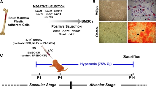
Objectives: Given the reported protective actions of bone marrow stromal cells (BMSCs; mesenchymal stem cells) in models of lung and cardiovascular injury, we tested their therapeutic potential in a murine model of BPD.

Methods: Neonatal mice exposed to hyperoxia (75% O(2)) were injected intravenously on Day 4 with either BMSCs or BMSC-conditioned media (CM) and assessed on Day 14 for lung morphometry, vascular changes associated with pulmonary hypertension, and lung cytokine profile.

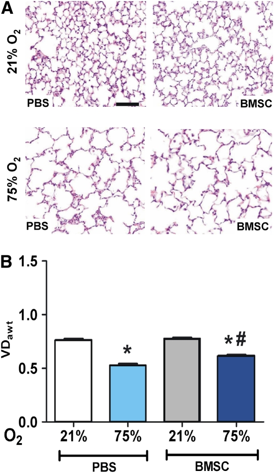
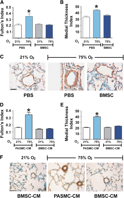
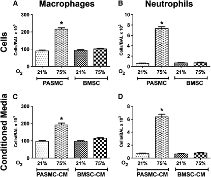
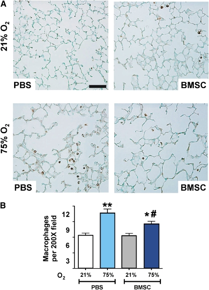
Measurements and main results: Injection of BMSCs but not pulmonary artery smooth muscle cells (PASMCs) reduced alveolar loss and lung inflammation, and prevented pulmonary hypertension. Although more donor BMSCs engrafted in hyperoxic lungs compared with normoxic controls, the overall low numbers suggest protective mechanisms other than direct tissue repair. Injection of BMSC-CM had a more pronounced effect than BMSCs, preventing both vessel remodeling and alveolar injury. Treated animals had normal alveolar numbers at Day 14 of hyperoxia and a drastically reduced lung neutrophil and macrophage accumulation compared with PASMC-CM-treated controls. Macrophage stimulating factor 1 and osteopontin, both present at high levels in BMSC-CM, may be involved in this immunomodulation.

Conclusions: BMSCs act in a paracrine manner via the release of immunomodulatory factors to ameliorate the parenchymal and vascular injury of BPD in vivo. Our study suggests that BMSCs and factor(s) they secrete offer new therapeutic approaches for lung diseases currently lacking effective treatment.


Figures

Figure 1.
Figure 1.
Isolation and differentiation of bone marrow stromal cell (BMSC) cultures and experimental design for in vivo study. (A) Cells were isolated from bone marrow and subjected to negative and positive selection using the indicated cell surface markers. (B) BMSCs were grown in the presence of adipogenic or osteogenic media, as described in Materials and Methods. After 3 weeks of adipogenic induction, lipid droplets could be observed within the cells (upper left panel), which were then stained using Oil Red O, as indicated by arrowheads (upper right panel). After 3 weeks of osteogenic induction (lower left panel), calcium deposition was visualized using Alizarin Red S staining, as indicated by arrows (lower right panel). (C) Neonatal mouse pups were exposed to hyperoxia on Postnatal Day 1, injected with cells or conditioned media on Postnatal Day 4, and killed on Day 14.
Figure 2.
Figure 2.
Effect of bone marrow stromal cell (BMSC) treatment on hyperoxic alveolar injury. (A) Representative hematoxylin and eosin–stained lung sections from normoxic, phosphate-buffered saline (PBS)- or BMSC-treated animals (upper panels) and from animals exposed to hyperoxia for 14 days and treated with PBS or BMSCs, as indicated on lower panels (original magnification ×100). Solid bar scale represents 400 μm and all the panels are under the same magnification. (B) Hyperoxia reduced the volume density of alveolar wall tissue (VDawt) compared with the normoxic group, indicative of lower alveolar count, which was modestly improved with BMSC treatment. Data are expressed as mean ± SEM (n = 10–12 animals per group). *P < 0.001 versus 21% O2 groups; #P < 0.01 versus PBS-treated hyperoxic group.
Figure 3.
Figure 3.
Either bone marrow stromal cell (BMSC) or BMSC–conditioned media (CM) treatment prevents vascular changes associated with pulmonary hypertension in hyperoxia-induced lung injury. (A) Hyperoxia-exposed, phosphate-buffered saline (PBS)-treated newborn mice develop significant right ventricular hypertrophy that is significantly reduced by BMSC treatment. Data are expressed as mean ± SEM (n = 10–12 animals per group). *P < 0.001, compared with the two normoxia groups and the hyperoxia group that received BMSC. (B) BMSC treatment significantly reduced the medial wall thickness as compared with the hyperoxia group that received PBS treatment. Data are expressed as mean ± SEM. *P < 0.001, compared with normoxia and the hyperoxia group that received BMSC. (C) Representative pulmonary arterioles immunostained for α–smooth muscle actin, displaying a thickened smooth muscle layer in hyperoxia-exposed mouse lungs as compared with normoxic controls, and absence of muscularization on BMSC treatment. (D) Similar to BMSC treatment, BMSC-CM treatment significantly reduced right ventricular hypertrophy in hyperoxia-exposed animals, and (E) significantly reduced medial wall thickness as compared with the hyperoxia group that received pulmonary artery smooth muscle cell–CM. Data are expressed as mean ± SEM (n = 16–18 animals per group). *P < 0.0001 versus normoxic groups or BMSC-CM treated groups. (F) Representative small pulmonary arterioles, as in (C). Solid bar scale represents 100 μm and all the panels are under the same magnification.
Figure 4.
Figure 4.
Effect of bone marrow stromal cell–conditioned media (BMSC-CM) on hyperoxic alveolar injury. (A) Representative hematoxylin and eosin–stained lung sections from normoxic animals compared with hyperoxic animals treated with either pulmonary artery smooth muscle cell (PASMC)-CM or BMSC-CM, as indicated. Quantitation of volume density of alveolar wall tissue (VDawt) is shown in (B). Treatment with BMSC-CM but not PASMC-CM prevented alveolar loss. Data are expressed as mean ± SEM (n = 16–18 animals per group). *P < 0.0001 versus normoxic controls and BMSC-CM group. Solid bar scale represents 400 μm and all the panels are under the same magnification.
Figure 5.
Figure 5.
Effect of stem cells or cell-free conditioned media (CM) treatments on bronchoalveolar lavage fluid (BALF) macrophage and neutrophil counts. (A and B) Differential cell counts for neutrophils and macrophages were performed in the BALF of normoxic or hyperoxic animals revealing marked inhibition of both cell types by treatment with bone marrow stromal cells (BMSCs) but not with pulmonary artery smooth muscle cells. Similarly, BMSC-CM suppressed both (C) macrophage, and (D) neutrophil numbers in bronchoalveolar lavage fluid of hyperoxic animals to levels of normoxic controls. Data are expressed as mean ± SEM (n = 16–18 animals per group). *P < 0.0001 versus normoxia or BMSC- or BMSC-CM–treated hyperoxic groups.
Figure 6.
Figure 6.
Effect of bone marrow stromal cell (BMSC) transplantation on lung inflammation. (A) Immunostaining with anti–Mac-3 antibody for macrophages (brown staining) in representative paraffin-embedded lung sections from BMSC recipient or phosphate-buffered saline (PBS) control mice under normoxic conditions (upper panels) or after 14 days of exposure to hyperoxia (lower panels). Original magnification: ×200. (B) Quantitation of Mac-3–positive cells. Hyperoxia-exposed lungs demonstrated a higher macrophage count compared with normoxia that was significantly reduced in the BMSC-treated hyperoxic group compared with the respective PBS control. Data are expressed as mean ± SEM (n = 6–8 animals per group) *P < 0.01, **P < 0.001 compared with normoxia groups; #P < 0.01, compared with the control hyperoxia group that received PBS. Solid bar scale represents 200 μm and all the panels are under the same magnification.

Comment in

References

    1. Kinsella JP, Greenough A, Abman SH. Bronchopulmonary dysplasia. Lancet 2006;367:1421–1431. - PubMed
    1. Stenmark KR, Abman SH. Lung vascular development: implications for the pathogenesis of bronchopulmonary dysplasia. Annu Rev Physiol 2005;67:623–661. - PubMed
    1. Abman SH. Pulmonary hypertension in chronic lung disease of infancy. Pathogenesis, pathophysiology and treatment. In: Bland RD, Coalson JJ, editors. Chronic lung disease of infancy. New York: Marcel Dekker; 2000. pp. 619–668.
    1. Baveja R, Christou H. Pharmacological strategies in the prevention and management of bronchopulmonary dysplasia. Semin Perinatol 2006;30:209–218. - PubMed
    1. Pereira RF, O'Hara MD, Laptev AV, Halford KW, Pollard MD, Class R, Simon D, Livezey K, Prockop DJ. Marrow stromal cells as a source of progenitor cells for nonhematopoietic tissues in transgenic mice with a phenotype of osteogenesis imperfecta. Proc Natl Acad Sci USA 1998;95:1142–1147. - PMC - PubMed

Publication types

MeSH terms