. 2009 Dec 3;114(24):4989-97.
doi: 10.1182/blood-2009-06-229567. Epub 2009 Aug 27.

B-cell follicle development remodels the conduit system and allows soluble antigen delivery to follicular dendritic cells

Affiliations

B-cell follicle development remodels the conduit system and allows soluble antigen delivery to follicular dendritic cells

Marc Bajénoff et al. Blood. .

Abstract

Afferent lymph is transported throughout lymph nodes (LNs) by the conduit system. Whereas this conduit network is dense in the T-cell zone, it is sparse in B-cell follicles. In this study, we show that this differential organization emerges during lymph node development. Neonatal LNs lack B follicles, but have a developed T-cell zone and a dense conduit network. As new T and B cells enter the developing LN, the conduit network density is maintained in the T, but not the B zone, leading to a profound remodeling of the follicular network that nevertheless maintains its connectivity. In adults, the residual follicular conduits transport soluble antigen to deep regions, where follicular dendritic cells are abundant and appear to replace the fibroblastic reticular cells that enwrap conduits in the T zone. This strategic location correlates with the capacity of the follicular dendritic cells to capture antigen even in the absence of antigen-specific antibodies. Together, these results describe how the stromal organization of the T and B regions of LNs diverges during development, giving rise to distinct antigen transport and delivery modes in the 2 compartments.


Figures

Figure 1
Figure 1
The conduit system of B-cell follicles is sparse and poorly branched. Peripheral LNs (except mesenteric) were collected from WT mice and cut in 40-μm-thick sections. LN sections were stained for B220 (light blue), CD3 (deep blue), ERTR-7 (red), and collagen IV (green) expression to reveal the complex structure of the conduit network present in the T- and B-cell zone when analyzed by confocal microscopy. Representative LN sections show the differences of the conduit network in the B- and the T-cell zones, as highlighted in the insets showing enlarged views of the conduits in the 2 areas. Data were acquired using a Leica Sp5 microscope (×20 and ×63 objectives) and are representative of 3 different experiments. Scale bar: 100 μm.
Figure 2
Figure 2
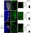
The conduit network is remodeled within developing B follicles. B cell–deficient (μMT) mice were injected with 2 × 10⁷ CMFDA-labeled WT B cells. Peripheral LNs (except mesenteric) were collected from WT mice (A) and μMT mice 1 day (B) and 2 weeks (C) after transfer. Sections were stained for B220 (white), CD3 (blue), ERTR-7 (green), and Lyve-1 (red) to reveal the status of the conduit network present in the T- and B-cell zone when analyzed by confocal microscopy. Histograms indicate the percentage of B-cell areas and T-cell areas occupied by the reticular fibers in each condition. Data were acquired using a Leica Sp5 microscope (×20 and ×63 objectives) and are representative of 3 different experiments. Scale bar: 100 μm.
Figure 3
Figure 3
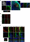
Follicle development is associated with conduit remodeling in neonates and allows FDCs to wrap around conduits. (A) WT mice were killed at day 3, 6, or 21 after birth. Axillary and inguinal LNs were harvested, sectioned, and stained for B220 (white), CD3 (blue), collagen IV (green), and FDC-M2 (red) to analyze by confocal microscopy the status of the conduit system in the developing follicles over time. Insets show enlargements of B- and T-cell areas. (B-C) LNs from adult irradiated ubiquitin-GFP mice reconstituted with WT bone marrow were sectioned and stained for collagen IV (red) and FDC-M2 (blue) expression to distinguish by confocal microscopy the very thin processes of the radioresistant GFP-positive (green)–expressing cells present in the follicles. Insets in panel C show an enlargement of a conduit and its enwrapping FDCs in the follicle. Data were acquired using a Leica Sp5 microscope (×20 and ×63 objectives) and are representative of 3 different experiments. Scale bar: 100 μm.
Figure 4
Figure 4
FDCs rapidly capture a soluble Ag injected subcutaneously. (A) WT mice were injected with 10 μL (50 μg) of Alexa 488–conjugated WGA (green) in the ears. Thirty minutes later, ear-draining LNs were collected; sectioned; stained for B220 (white), FDC-M2 (red), and collagen IV (blue) expression; and analyzed by confocal microscopy. Insets show the enlargement of a region that contains FDCs and conduits. (B) WT mice were injected with 10 μL (20 μg) of OVA or HEL in the ears. Ear-draining LNs were harvested 30 minutes later, sectioned, stained for HEL (green) and FDC-M2 (red) expression, and analyzed by confocal microscopy. (C) WT mice were injected with 10 μL (50 μg) of Alexa 488–conjugated WGA (green) in the ears. One, 5, 10, and 20 minutes later, ear-draining LNs were collected, sectioned, and stained for FDC-M2 (blue) and collagen IV (red) expression to examine the WGA diffusion over time when analyzed by confocal microscopy. Data were acquired with a Leica Sp5 microcope (×20 and ×63 objectives) and are representative of 3 different experiments. Scale bar: 100 μm.
Figure 5
Figure 5
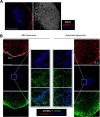
SCS macrophages are not responsible for Ag transport to FDCs. (A) LN sections from WT mice were stained for B220 (white), FDC-M2 (blue), and MOMA-1 (red), then analyzed by confocal microscopy to highlight the SCS macrophages and their dendritic protrusions that penetrate the follicles. (B) WT mice were injected with 30 μL of PBS- or chlodornate-loaded liposomes in the hind footpads. Six days later, when SCS macrophage depletion was complete, 10 μL (50 μg) of Alexa 647–conjugated WGA was injected in the footpads of the treated animals. Popliteal LNs were harvested 30 minutes later; sectioned; stained for MOMA-1 (white), collagen IV (red), and FDC-M2 (blue); and then analyzed by confocal microscopy. Data were acquired with a Leica Sp5 microcope (×20 and ×63 objectives) and are representative of 2 different experiments. Scale bar: 100 μm.

References

    1. Willard-Mack CL. Normal structure, function, and histology of lymph nodes. Toxicol Pathol. 2006;34:409–424. - PubMed
    1. Farr AG, Cho Y, De Bruyn PP. The structure of the sinus wall of the lymph node relative to its endocytic properties and transmural cell passage. Am J Anat. 1980;157:265–284. - PubMed
    1. Racz P, Tenner-Racz K, Myrvik QN, Ockers JR. The “mosaic” structure of the sinuses in the hilar lymph node complex of the lungs in rabbits undergoing a pulmonary cell-mediated reaction: an electron microscopic study. J Reticuloendothel Soc. 1978;24:527–545. - PubMed
    1. Clark SL., Jr The reticulum of lymph nodes in mice studied with the electron microscope. Am J Anat. 1962;110:217–257. - PubMed
    1. van Ewijk W, Brekelmans PJ, Jacobs R, Wisse E. Lymphoid microenvironments in the thymus and lymph node. Scanning Microsc. 1988;2:2129–2140. - PubMed

Publication types