Review
. 2009 Aug 28;105(5):408-21.
doi: 10.1161/CIRCRESAHA.109.201566.

Heart valve development: regulatory networks in development and disease

Affiliations
Review

Heart valve development: regulatory networks in development and disease

Michelle D Combs et al. Circ Res. .

Abstract

In recent years, significant advances have been made in the definition of regulatory pathways that control normal and abnormal cardiac valve development. Here, we review the cellular and molecular mechanisms underlying the early development of valve progenitors and establishment of normal valve structure and function. Regulatory hierarchies consisting of a variety of signaling pathways, transcription factors, and downstream structural genes are conserved during vertebrate valvulogenesis. Complex intersecting regulatory pathways are required for endocardial cushion formation, valve progenitor cell proliferation, valve cell lineage development, and establishment of extracellular matrix compartments in the stratified valve leaflets. There is increasing evidence that the regulatory mechanisms governing normal valve development also contribute to human valve pathology. In addition, congenital valve malformations are predominant among diseased valves replaced late in life. The understanding of valve developmental mechanisms has important implications in the diagnosis and management of congenital and adult valve disease.


Figures

Figure 1
Figure 1. Stratified ECM compartments are evident in mature SL and AV valves
A) Schematic representation of one of three valve cusps of the aortic or pulmonic SL valve with fibrosa (F), spongiosa (S) and ventricularis (V) layers indicated. B) Schematic representation of one AV valve leaflet with atrialis (A), spongiosa (S), and fibrosa (F) layers indicated. The mitral valve has two leaflets, whereas the tricuspid valve has three leaflets, all of which are supported by chordae tendineae (CT). The direction of pulsatile blood flow is indicated for both SL and AV valves (arrow).
Figure 2
Figure 2. Model for regulatory interactions that control endocardial cushion formation (A) and EMT (B)
A) Myocardial BMP2 expression increases hyaluronan and versican deposition in cushion-forming regions of the AVC and OFT. BMP2 induces Tbx2 transcription in the myocardium, inhibiting chamber-specific gene expression. VEGF, expressed in endothelial cells, promotes endocardial cushion endothelial cell proliferation. B) Myocardial expression of BMP2 promotes endocardial cushion EMT. Multiple endocardially-derived signals promote endocardial cushion EMT (delaminating and mesenchymal cells are indicated by white stars). TGFβ signals through Slug to promote EMT, while Notch1 signals through Snail to suppress VE-cadherin (VE-cad) expression and promote EMT. Wnt/β-catenin signaling increases endocardial cushion EMT. Once the cushions are established, endocardial VEGF expression maintains endothelial cell proliferation and inhibits EMT.
Figure 3
Figure 3. Model for regulatory interactions that control growth of endocardial cushions/valve primordia
Cell proliferation in the endothelial cells in the endocardial cushions is induced by VEGF/NFATc1 and Shp2/ERK1/2 signaling. Mesenchymal cell proliferation is induced by multiple signaling mechanisms including Wnt/β-catenin, TGFβs, BMPs, FGF4 and Shp2/ERK1/2. EGF signaling inhibits mesenchymal cell proliferation.
Figure 4
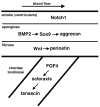
Figure 4. Model for regulatory interactions controlling AV valve stratification and lineage diversification
Notch1 expression is localized to the flow side of the stratifying valve. In the spongiosa, BMP2 signaling promotes Sox9 expression and deposition of cartilage-related ECM components, such as aggrecan. Wnt signaling in the fibrosa promotes expression of fibroblast/pre-osteoblast-related ECM components, such as periostin. Maturation of valve supporting structures (chordae tendineae) is associated with FGF4 signaling, which induces expression of the tendon-related transcription factor scleraxis and the ECM component tenascin. Although the SL valves do not have chordae tendineae, these signaling pathways also are active in the corresponding regions of the stratified aortic valve cusps and supporting structures.

References

    1. Hoffman JIE, Kaplan S. The incidence of congenital heart disease. J Am Coll Cardiol. 2002;39:1890–1900. - PubMed
    1. Pierpont ME, Basson CT, Benson DW, Jr, Gelb BD, Giglia TM, Goldmuntz E, McGee G, Sable CA, Srivastava D, Webb CL. Genetic basis for congenital heart defects: current knowledge: a scientific statement from the American Heart Association Congenital Cardiac Defects Committee, Council on Cardiovascular Disease in the Young. Circulation. 2007;115:3015–3038. - PubMed
    1. Supino PG, Borer JS, Yin A, Dillingham E, McClymont W. The epidemiology of valvular heart diseases: the problem is growing. Adv Cardiol. 2004;41:9–15. - PubMed
    1. Roberts WC, Ko JM. Frequency by decades of unicuspid, bicuspid and tricuspid aortic valves in adults having isolated aortic valve replacement for aortic stenosis, with or without associated aortic regurgitation. Circulation. 2005;111:920–925. - PubMed
    1. Weismann CG, Gelb BD. The genetics of congenital heart disease: a review of recent developments. Curr Opin Cardiol. 2007;22:200–206. - PubMed

Publication types

MeSH terms