. 2009 Oct 21;28(20):3196-206.
doi: 10.1038/emboj.2009.246. Epub 2009 Aug 27.

Wip1 confers G2 checkpoint recovery competence by counteracting p53-dependent transcriptional repression

Affiliations

Wip1 confers G2 checkpoint recovery competence by counteracting p53-dependent transcriptional repression

Arne Lindqvist et al. EMBO J. .

Abstract

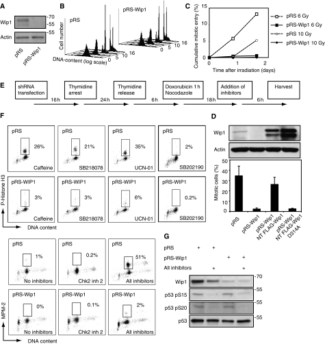
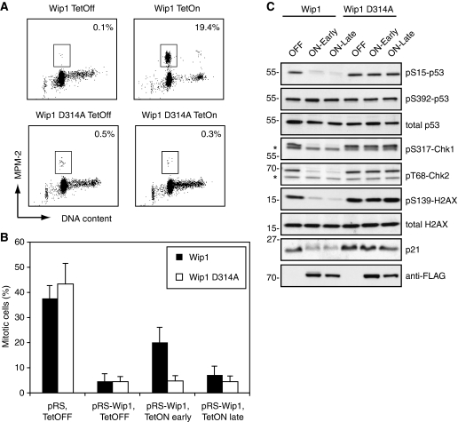
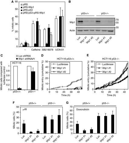
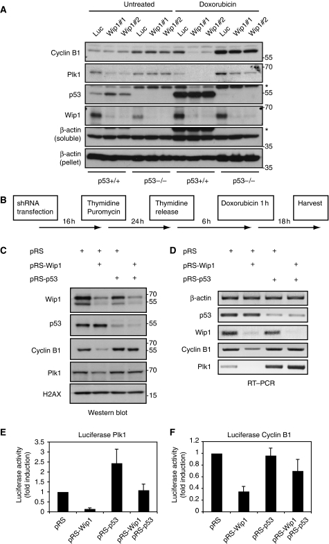
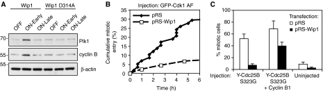
Activation of the DNA damage checkpoint causes a cell-cycle arrest through inhibition of cyclin-dependent kinases (cdks). To successfully recover from the arrest, a cell should somehow be maintained in its proper cell-cycle phase. This problem is particularly eminent when a cell arrests in G2, as cdk activity is important to establish a G2 state. Here, we identify the phosphatase Wip1 (PPM1D) as a factor that maintains a cell competent for cell-cycle re-entry during an ongoing DNA damage response in G2. We show that Wip1 function is required throughout the arrest, and that Wip1 acts by antagonizing p53-dependent repression of crucial mitotic inducers, such as Cyclin B and Plk1. Our data show that the primary function of Wip1 is to retain cellular competence to divide, rather than to silence the checkpoint to promote recovery. Our findings uncover Wip1 as a first in class recovery competence gene, and suggest that the principal function of Wip1 in cellular transformation is to retain proliferative capacity in the face of oncogene-induced stress.


Conflict of interest statement

The authors declare that they have no conflict of interest.

Figures

Figure 1
Figure 1
Wip1 is essential for checkpoint recovery. (A) Wip1 RNAi efficiently reduces the levels of endogenous Wip1. U2OS cells, transfected with empty pRS or pRS-Wip1, were selected with puromycin and subjected to immunoblotting with the indicated antibodies. (B) Wip1 RNAi cells progress through S, G2 and M-phases in the absence of DNA-damage. U2OS cells co-transfected with GFP-Spectrin and pRS-Wip1 or empty pRS were synchronized in S-phase with a single 24 h thymidine block. The cell-cycle profile of GFP-expressing cells was assessed by FACS at different time-points after release. Y-axis: number of cells, x-axis: DNA-content (logarithmic scale), z-axis: time after thymidine release (hours). (C) Wip1 RNAi inhibits checkpoint recovery. Cells were treated as in (B), but subjected to 6 or 10 Gy γ-irradiation 7 h after release from the thymidine block. Immediately after irradiation or after 24 h incubation, taxol was added for 24 h before harvesting. The amount of mitotic GFP-positive cells was assessed by MPM-2 positivity using FACS. (D) Wip1 RNAi inhibits caffeine-induced checkpoint recovery. U2OS cells were co-transfected with GFP-spectrin and pRS, pRS-Wip1, pRS-Wip1 + RNAi insensitive FLAG-Wip1, or pRS-Wip1 + RNAi-insensitive phosphatase dead FLAG-Wip1 (D314A) and synchronized by a single 24 h thymidine block. Six hours after thymidine synchronization, cells were treated with the topoisomeraseII inhibitor doxorubicin, and nocodazole was added to trap cells entering mitosis. After 15 h, cells were treated for 6 h with caffeine (inhibitor of ATM/ATR) and scored for phosphorylated Histone H3 by FACS. Graph denotes average of three independent experiments, error bars represent standard error. Western blot shows endogenous and reconstituted Wip1 levels from a single experiment. (E) Outline of experimental setup used in (F, G) and Figure 3A. (F) A range of kinase inhibitors does not promote checkpoint recovery after Wip1 depletion. Cells were treated as in (B), but treated with a 1-h pulse of doxorubicin 6 h after release from thymidine block. After 18 h, cells were treated for 6 h with caffeine (inhibitor of ATM/ATR), SB218078 (inhibitor of Chk1), UCN-01 (inhibitor of Chk1 and MK-2), SB202190 (inhibitor of p38), Chk2 inhibitor-2 (Chk2 inhibitor), or a combination of these inhibitors. The percentage of mitotic cells was assessed by scoring GFP positive cells for P-Histone H3 or MPM2 positivity by FACS. (G) P53 is dephosphorylated on the ATM/ATR-site S15 and the Chk1/Chk2 site S20 after combined inhibitor treatment (Shieh et al, 2000; Appella and Anderson, 2001). Cells treated or not with all inhibitors as in Figure 1F were subjected to western blot with the indicated antibodies.
Figure 2
Figure 2
Wip1 is required throughout the checkpoint arrest to mediate recovery. (A) Forced expression of Wip1 can induce checkpoint recovery. U2TR cells stably expressing inducible wt or phosphatase-deficient Wip1 (D314A) were synchronized by thymidine and 6 h after release treated with doxorubicin for 1 h, after which the cells were treated for 24 h with Tetracyclin and taxol. The percentage of mitotic cells was assessed by scoring MPM2 positivity by FACS. (B) Wip1 is required during a G2 checkpoint arrest. U2TR cells stably expressing inducible RNAi-resistant wt or phosphatase-deficient Wip1 (D314A) were co-transfected with GFP-Spectrin and pRS or pRS-Wip1. Cells were synchronized with thymidine and 6 h after release treated with doxorubicin for 1 h, followed by taxol addition. Tetracyclin was added immediately (early) or 18 h (late) after doxorubicin washout. Caffeine was added 18 h after doxorubicin washout and incubated for 8 h. The percentage of mitotic cells was assessed by scoring GFP positive cells for MPM2 positivity by FACS. (C) Checkpoint signalling is inhibited both after early and late Wip1-induction. Cells were treated as in (B), but selected with puromycin and without caffeine addition, harvested as in (B) and subjected to immunoblotting with the indicated antibodies.
Figure 3
Figure 3
Wip1 is essential for checkpoint recovery by antagonizing p53. (A) p53 RNAi reverses Wip1 phenotype. U2OS cells were transfected with empty pRS, pRS + pRS-Wip1, pRS + pRS-p53 or pRS-Wip1 + pRS-p53. All samples were co-transfected with GFP-Spectrin. Checkpoint recovery was assessed as in Figure 1E and F. Graph denotes percentage of mitotic cells and error bars represent standard error of three independent experiments. (B) Knockdown of Wip1 in HCT116 cells. HCT116 p53+/+ or p53−/− cells were infected with lentiviral shRNA constructs targeting luciferase or Wip1. After selection with puromycin, harvested cells were subjected to immunoblotting with the indicated antibodies. (C) Wip1 is necessary for caffeine-induced checkpoint recovery in HCT116 cells, but not in HCT116 p53−/− cells. HCT116 and HCT116 p53−/− cells were infected with lentivirus expressing Wip1 shRNA#1 or luciferase shRNA, selected with puromycin and treated with doxorubicin for 16 h. Caffeine and taxol, to induce checkpoint recovery and to trap cells in mitosis, respectively, were added for 12 h after the doxorubicin treatment. Graph shows the ratio of mitotic Wip1 RNAi cells in relation to the amount of mitotic luciferase RNAi cells. (D, E) Wip1 is necessary for checkpoint recovery in HCT116 cells, but not in HCT116 p53−/− cells. HCT116 and HCT116 p53−/− cells were infected with lentivirus expressing Wip1 shRNA or luciferase shRNA and selected with puromycin. After treatment with 0.75 μM doxorubicin for 2 h, cells were followed by time-lapse microscopy and scored for entry into mitosis. (F, G) Wip1 is necessary for checkpoint recovery from late S/G2 in HCT116 cells, but not in HCT116 p53−/− cells. HCT116 and HCT116 p53−/− cells were infected with lentivirus expressing Wip1 shRNA or luciferase shRNA and selected with puromycin. Four hours after a BrdU-pulse, cells were treated with 6 Gy γ-irradiation or 1 h of 0.5 μM doxorubicin. Taxol was added immediately after DNA damage, and cells were harvested 24 h later. The percentage of recovering cells was assessed by scoring MPM2 positivity on the BrdU-positive population by FACS.
Figure 4
Figure 4
Wip1 counteracts p53-mediated transcriptional repression. (A) Wip1 antagonizes p53-mediated downregulation of Plk1 and Cyclin B1 in HCT116 cells. HCT116 and HCT116 p53−/− cells were infected with lentivirus expressing Wip1 shRNA or a luciferase shRNA, selected with puromycin, treated with doxorubicin for 16 h and harvested for western blot. Whereas Wip1 was detected in the unsoluble fraction (loading control, β-actin pellet), p53, Cyclin B1 and Plk1 were detected in the soluble fraction. (B) Schematic outline of experiments in (C–F). (C, D) Wip1 antagonizes p53-mediated downregulation of Plk1 and Cyclin B1. U2OS cells were transfected as in Figure 3A, and selected by puromycin addition during a 24-h thymidine block. At 6 h after release from thymidine, cells were treated for 1 h with doxorubicin. After 18 h, at the time corresponding to addition of inhibitors in Figure 3A, cells were harvested for western blot (C) or RT–PCR (D). (E, F) Wip1 counteracts p53-mediated transcriptional repression of Plk1 and Cyclin B1. U2OS cells were transfected as in Figure 3A, with the addition of luciferase-based transcriptional reporters for Plk1 (E) and Cyclin B1 (F), and synchronized as shown in Figure 4B. Graphs show the relative luciferase expression 18 h after doxorubicin treatment.
Figure 5
Figure 5
Cyclin B1 is rate limiting for recovery in Wip1-depleted cells. (A) Plk1 and Cyclin B1 levels are low if Wip1 is absent during the DNA damage arrest. The samples used in Figure 2C were subjected to immunoblot for Plk1 and Cyclin B1. (B) Injection of GFP-Cdk1 AF fails to induce mitotic entry in DNA-damaged Wip1-depleted cells. pRS or pRS-Wip1 transfected cells were selected with puromycin and 6 h after release from a thymidine block exposed for 1 h to 0.5 μM doxorubicin. At 18 h after doxorubicin treatment, GFP-Cdk1 AF was injected and cells were followed by time-lapse microscopy. Graph denotes cumulative mitotic entry of injected cells (C) Injection of YFP-Cdc25B3 S323G fails to induce mitotic entry in DNA-damaged Wip1 RNAi cells, unless co-injected with Cyclin B1. pRS or pRS-Wip1 transfected cells were selected with puromycin and 6 h after release from a thymidine block exposed for 1 h to 0.5 μM doxorubicin. At 18 h after doxorubicin treatment, YFP-Cdc25B3 S323G or YFP-Cdc25B3 S323G and Cyclin B1 were injected to cells in a single dish and nocodazole was added to trap cells entering mitosis. Graph denotes average of four independent experiments where 23–178 cells were counted for each injected construct, error bars represent standard error.

References

    1. Appella E, Anderson CW (2001) Post-translational modifications and activation of p53 by genotoxic stresses. Eur J Biochem 268: 2764–2772 - PubMed
    1. Banin S, Moyal L, Shieh S, Taya Y, Anderson CW, Chessa L, Smorodinsky NI, Prives C, Reiss Y, Shiloh Y, Ziv Y (1998) Enhanced phosphorylation of p53 by ATM in response to DNA damage. Science 281: 1674–1677 - PubMed
    1. Bassermann F, Frescas D, Guardavaccaro D, Busino L, Peschiaroli A, Pagano M (2008) The Cdc14B-Cdh1-Plk1 axis controls the G2 DNA-damage-response checkpoint. Cell 134: 256–267 - PMC - PubMed
    1. Bartkova J, Rezaei N, Liontos M, Karakaidos P, Kletsas D, Issaeva N, Vassiliou LVF, Kolettas E, Niforou K, Zoumpourlis VC, Takaoka M, Nakagawa H, Tort F, Fugger K, Johansson F, Sehested M, Andersen CL, Dyrskjot L, Orntoft T, Lukas J et al. (2006) Oncogene-induced senescence is part of the tumorigenesis barrier imposed by DNA damage checkpoints. Nature 444: 633–637 - PubMed
    1. Batchelor E, Mock CS, Bhan I, Loewer A, Lahav G (2008) Recurrent initiation: a mechanism for triggering p53 pulses in response to DNA damage. Mol Cell 30: 277–289 - PMC - PubMed

Publication types

MeSH terms