. 2009 Aug;5(8):e1000562.
doi: 10.1371/journal.ppat.1000562. Epub 2009 Aug 28.

Regulation of host translational machinery by African swine fever virus

Affiliations

Regulation of host translational machinery by African swine fever virus

Alfredo Castelló et al. PLoS Pathog. 2009 Aug.

Abstract

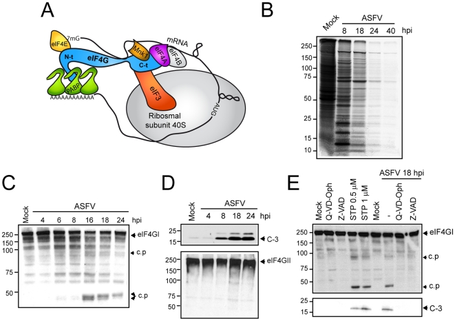
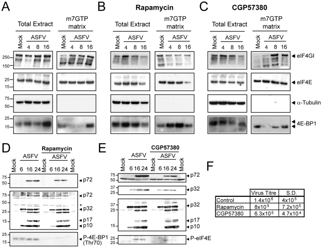
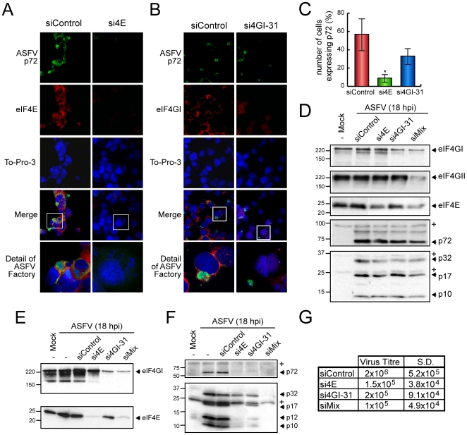
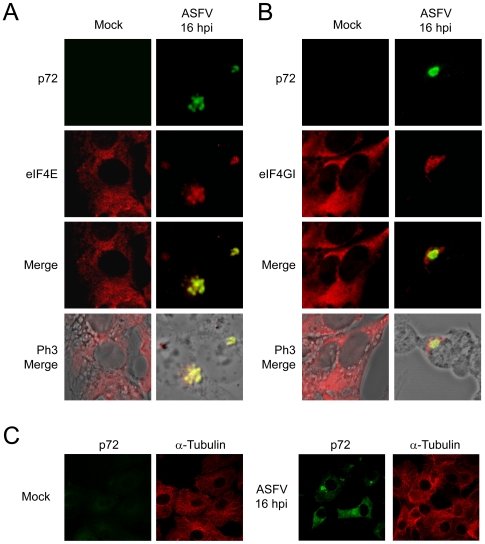
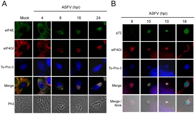
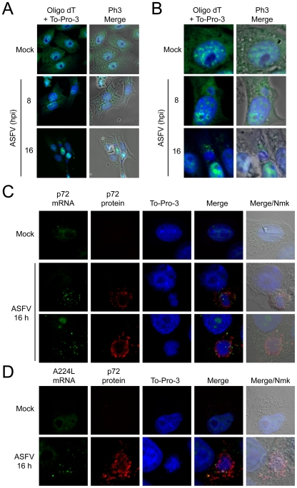
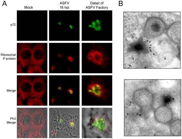
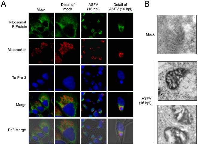
African swine fever virus (ASFV), like other complex DNA viruses, deploys a variety of strategies to evade the host's defence systems, such as inflammatory and immune responses and cell death. Here, we analyse the modifications in the translational machinery induced by ASFV. During ASFV infection, eIF4G and eIF4E are phosphorylated (Ser1108 and Ser209, respectively), whereas 4E-BP1 is hyperphosphorylated at early times post infection and hypophosphorylated after 18 h. Indeed, a potent increase in eIF4F assembly is observed in ASFV-infected cells, which is prevented by rapamycin treatment. Phosphorylation of eIF4E, eIF4GI and 4E-BP1 is important to enhance viral protein production, but is not essential for ASFV infection as observed in rapamycin- or CGP57380-treated cells. Nevertheless, eIF4F components are indispensable for ASFV protein synthesis and virus spread, since eIF4E or eIF4G depletion in COS-7 or Vero cells strongly prevents accumulation of viral proteins and decreases virus titre. In addition, eIF4F is not only activated but also redistributed within the viral factories at early times of infection, while eIF4G and eIF4E are surrounding these areas at late times. In fact, other components of translational machinery such as eIF2alpha, eIF3b, eIF4E, eEF2 and ribosomal P protein are enriched in areas surrounding ASFV factories. Notably, the mitochondrial network is polarized in ASFV-infected cells co-localizing with ribosomes. Thus, translation and ATP synthesis seem to be coupled and compartmentalized at the periphery of viral factories. At later times after ASFV infection, polyadenylated mRNAs disappear from the cytoplasm of Vero cells, except within the viral factories. The distribution of these pools of mRNAs is similar to the localization of viral late mRNAs. Therefore, degradation of cellular polyadenylated mRNAs and recruitment of the translation machinery to viral factories may contribute to the inhibition of host protein synthesis, facilitating ASFV protein production in infected cells.


Conflict of interest statement

The authors have declared that no competing interests exist.

Figures

Figure 1
Figure 1. Inhibition of host protein synthesis and analysis of caspase-3-mediated eIF4GI cleavage in ASFV-infected cells.
A) Schematic representation of translation initiation complex. B) Cellular and viral protein synthesis during ASFV infection. Cultures of Vero cells (5×10⁵) were mock infected (Mock) or infected with ASFV (5 pfu/cell), and labeled at different times after infection with 200 µCi of [35S]Met-[35S]Cys/ml in cysteine-methionine-free medium for 2 h. Samples were analyzed by SDS-PAGE followed by fluorography and autoradiography. C) Activation of caspase-3 during ASFV infection induces incomplete eIF4GI degradation. eIF4GI was detected by Western blot by incubation with specific antiserum at the indicated times after ASFV-infected Vero cells. D) eIF4GII (upper panel) and cleaved caspase-3 (bottom panel) were analyzed by Western blotting. E) Analysis of eIF4G1 cleavage by using specific caspase-3 inducers or inhibitors in mock-infected or ASFV-infected cells. Vero cells were treated with 0.5 or 1 µM staurosporin or were infected with ASFV (5 pfu/cell). Two replicates of mock-infected or ASFV-infected cells were treated with 60 µM Q-VD-Oph or Z-VAD. Cells were recovered after 18 h in sample buffer and eIF4GI (upper panel) and caspase-3 (bottom panel) activation was analyzed by Western blot. c.p., cleavage product; C-3, cleaved caspase-3 (17 KDa); STP, staurosporin.
Figure 2
Figure 2. Effect of ASFV infection on total level and phosphorylation status of eIF2α, eIF4E, eIF4GI and 4E-BP1.
A) Steady-state levels of eIF2α, phospho-eIF2α and PKR. At the indicated times after ASFV infection, Vero cells (MOI = 5 pfu/cell) were solubilised in sample buffer and equivalent amounts of protein were analyzed by Western blot with specific antisera. B) Phosphorylation of Mnk-1 and eIF4E is stimulated in ASFV-infected Vero cells. Vero cells were either mock-infected (Mock) or infected with ASFV (MOI = 5 pfu/cell). At the indicated times (hpi), total protein was isolated, and equivalent amounts were fractionated by SDS-PAGE, and analyzed by immunoblotting using antisera recognizing phospho-eIF4E (P-eIF4E), total eIF4E, phospho Mnk-1 (P-Mnk-1), eIF4A and eEF2. C) The phosphorylation status of 4E-BP1 was analyzed using antibodies against total 4E-BP1 (upper panel), phopho-4E-BP1 (Thr70) (middle panel) and non-phospho-4E-BP1 (Thr46) (bottom panel). D) ASFV infection increases level of phosphorylated eIF4G through mTOR activation. Vero cells were non-treated or pretreated for 12 h with rapamycin 250 nM and then infected with 5 pfu/cell of ASFV. Cells were then lysed with buffer sample at 4, 8 and 18 hpi. Amounts of phospho-eIF4GI and total eIF4GI were analyzed by Western blot (upper panels). The phosphorylation status of Mnk-1 in the absence or presence of rapamycin was analyzed by using specific antiserum anti-phospho-Mnk-1 (middle-bottom panel). α-Tubulin was detected as a load control (bottom panel). E) The ASFV-induced phosphorylation of eIF4G and Mnk-1 requires late events of the viral cycle. ASFV-infected Vero cells were treated with AraC (40 µg/ml) throughout the infection course. Cells were then lysed and 30 µg of protein were subjected to electrophoresis and analyzed by Western blot with specific antisera against eIF4GI, phospho-eIF4GI (upper panels) and phospho-Mnk-1 (middle-bottom panel). α-Tubulin was detected with a specific antibody as a load control (bottom panel).
Figure 3
Figure 3. Analysis of cap-binding complex during ASFV-infection.
A) Interaction of eIF4GI with eIF4E is increased upon ASFV infection. Vero cells were infected with 5 pfu/cell of ASFV and lysed at 4, 8 or 16 hpi with buffer A. Cell extracts were then incubated with Sepharose 4B matrix followed by Sepharose-4B-m7GTP matrix. Cap-binding complexes were eluted with Laemmli sample buffer. eIF4E, eIF4GI, α-Tubulin and 4E-BP1 were detected in total and eluted fractions by Western blotting. B and C) eIF4GI-eIF4E association is abrogated by rapamycin but not by CGP57380. Vero cells were pretreated with 250 nM rapamycin (B) or 20 µM CGP57380 (C). After 12 h cells were infected with ASFV (5 pfu/cell) in the continuous presence of the compounds. At 4, 8 and 16 hpi cells were recovered in buffer A and then incubated with Sepharose 4B matrix followed by Sepharose-4B-m7GTP matrix. eIF4GI, eIF4E, α-Tubulin and 4E-BP1 were detected in total and eluted fractions by Western blotting. D, E and F) Rapamycin and CGP57380 treatments only partially inhibit ASFV protein synthesis and virus spread. Vero cells were either pretreated or not with 250 nM rapamycin (D) or 20 µM CGP57380 (E). After 12 h cells were infected with ASFV (5 pfu/cell) in the continuous presence of the compounds. At 6, 16 and 24 hpi cells were recovered and viral proteins were analyzed with p72 (D and E, upper panel) and p32 (E, upper-middle panel) antibodies or an antisera that recognize most of the ASFV structural proteins (D and E middle-bottom panel). Phosphorylation of 4E-BP1 or eIF4E was analyzed with phosphospecific antibodies against Thr70 (D, bottom panel) and Ser209 (E, bottom panel), respectively. F) After 48 h of ASFV infection, supernatants from rapamycin or CGP57380 treated or untreated cells were recovered. Lytic viruses were titrated in Vero monolayers and plotted in the table. S.D., standard deviations.
Figure 4
Figure 4. Depletion of eIF4E or/and eIF4GI blocks ASFV proteins expression in infected cells.
A, B and C) COS-7 cells were transfected with siControl, si4E or si4GI-31 in 2 steps separated by 24 hours. Cells were then seeded on glass coverslips and mock infected or infected with 1 pfu/cell of ASFV. At 16 hpi, cells were permeabilised and fixed and ASFV p72 and eIF4E or eIF4GI were detected by indirect immunofluorescence. A and B) Immunofluorescence using anti-eIF4E or anti-eIF4GI, respectively, and anti-p72 in either eIF4E (A) or eIF4GI (B) silenced cells. C) Percentage of cells expressing ASFV p72 in eIF4E- and eIF4GI-silenced cells (mean±SD). * P<0.05. D) COS-7 cells were transfected with siControl, si4GI-31, si4E or a mixture of si4GI-31, si4E and si4GII-2 (siMix) as indicated. After 72 h, cells were infected with 1 pfu/cell of ASFV and samples were recovered after 18 hpi. Depletion of eIFs was examined by Western blot against eIF4GI, eIF4GII and eIF4E (upper panels). Accumulation of viral proteins was analyzed using a specific antibody against p72 (middle bottom panel) or with an antiserum that recognizes a number of structural ASFV proteins (bottom panel). E, F and G) Vero cells were transfected with siControl, si4GI-31, si4E or a mixture of si4GI-31 and si4E (siMix) as described. After 72 h, cells were infected with 1 pfu/cell of ASFV and samples were recovered after 18 hpi. Depletion of eIFs was examined by Western blot against eIF4GI and eIF4E (E). Accumulation of viral proteins was analyzed using a specific antibody against p72 (F, upper panel) or with an antiserum that recognizes a number of structural ASFV proteins (F, bottom panel). G) In parallel, supernatants from transfected cells were recovered at 48 hpi and titrated in Vero cells. Virus titre in each case was indicated in the table. * unspecific cellular protein detected by the antibody.
Figure 5
Figure 5. eIF4E and eIF4GI are recruited to ASFV factories in infected cells.
Vero cells were seeded on glass coverslips and mock infected or infected with 5 pfu/cell of ASFV. At 16 hpi cells were permeabilized, fixed and then eIF4E (A) or eIF4GI (B) and ASFV p72 were detected by indirect immunofluorescence. Cells were visualized by confocal microscopy and the cell outline was defined by phase contrast microscopy. C) In parallel, α-Tubulin and p72 were detected in infected (right panels) and mock cells (left panels) with specific antibodies. Images were obtained under restricted conditions and processed with Huygens 3.0 software.
Figure 6
Figure 6. eIF4E and eIF4GI redistribution during ASFV time-infection steps.
A) Vero cells were seeded on glass coverslips and mock infected or infected with 5 pfu/cell of ASFV. Cells were then permeabilized and fixed at 4, 8, 16 and 24 hpi. eIF4E and eIF4GI were detected by indirect immunofluorescence and cell nuclei and ASFV factories were stained with To-Pro-3. Cells were visualized by confocal microscopy and the cell outline was defined by phase contrast microscopy. Images were obtained under restricted conditions and processed with Huygens 3.0 software. B) Vero cells were seeded on glass coverslips and mock infected or infected with 5 pfu/cell of ASFV. Cells were then permeabilized and fixed at 8, 10 and 18 hpi. eIF4GI and p72 were detected by indirect immunofluorescence and cell nuclei and ASFV factories were stained with To-Pro-3. Cells were visualized by confocal microscopy and the cell outline was defined by nomarski microscopy.
Figure 7
Figure 7. Localization of mRNAs in ASFV-infected Vero cells.
Vero cells were seeded on glass coverslips and mock infected or infected with 5 pfu/cell of ASFV. At 8 and 16 hpi cells were fixed and permeabilized. Then, in situ hybridization with fluorescein labeled probes was carried out for each post-infection time. A) Distribution of polyadenylated mRNAs bulk by using oligo d(T) fluoresecein labelled probe. To-Pro 3 was used to stain cell nuclei and ASFV factories. B) Detail of mock infected and ASFV-infected cells, showing the polyadenylated mRNAs surrounding the viral factories. In situ hybridization with fluorescein labeled p72 (C) and A224L (D) probes. To-Pro 3 and p72 antibody were used to detect cell nuclei and ASFV factories. Cells were visualized by confocal microscopy and the cell outline was defined by phase contrast microscopy. Images were processed with Huygens 3.0 software.
Figure 8
Figure 8. P ribosomal protein is clustered surrounding ASFV factories.
A) Vero cells were seeded on glass coverslips and mock infected or infected with 5 pfu/cell of ASFV. At 16 hpi cells were permeabilized, fixed and ribosomal P protein and ASFV p72 were detected by indirect immunofluorescence by using specific antisera. Cells were visualized by confocal microscopy and the cell outline was defined by phase contrast microscopy. Images were then processed with Huygens 3.0 software. B) Electron microscopy detection of ribosomes in ASFV-assembly areas. Cells were infected with 5 pfu/cell of ASFV and processed for electron microscopy at 16 hpi. Ribosomes were detected using a specific monoclonal antibody against P ribosomal protein.
Figure 9
Figure 9. P ribosomal protein and mitochondrial network co-localize surrounding ASFV factories.
A) Vero cells were seeded on glass coverslips and infected with 5 pfu/cell of ASFV. For mitochondrial staining, cells were incubated at 15 hpi with 2 µM MitoTracker red CMH2-Ros for 45 min and then permeabilized and fixed. Ribosomal P protein was detected by indirect immunofluorescence and cell nuclei and viral factories were stained with To-Pro-3. Cells were visualized by confocal microscopy and the cell outline was defined by phase contrast microscopy. Images were obtained under restricted conditions and processed with Huygens 3.0 software. B) Detection by electron microscopy of ribosomes in mitochondria-containing areas. Cells were mock infected or infected with 5 pfu/cell of ASFV and processed for electron microscopy at 16 hpi. Ribosomes were detected using a specific monoclonal antibody against P ribosomal protein.

References

    1. Bushell M, Sarnow P. Hijacking the translation apparatus by RNA viruses. J Cell Biol. 2002;158:395–399. - PMC - PubMed
    1. Mohr I. Phosphorylation and dephosphorylation events that regulate viral mRNA translation. Virus Res. 2006;119:89–99. - PubMed
    1. Schneider RJ, Mohr I. Translation initiation and viral tricks. Trends Biochem Sci. 2003;28:130–136. - PubMed
    1. Prevot D, Darlix JL, Ohlmann T. Conducting the initiation of protein synthesis: the role of eIF4G. Biol Cell. 2003;95:141–156. - PubMed
    1. Imataka H, Gradi A, Sonenberg N. A newly identified N-terminal amino acid sequence of human eIF4G binds poly(A)-binding protein and functions in poly(A)-dependent translation. Embo J. 1998;17:7480–7489. - PMC - PubMed

Publication types