Review
. 2009 Jul-Aug;14(4):275-82.
doi: 10.1155/2009/380384.

Herpes zoster and postherpetic neuralgia: past, present and future

Affiliations
Review

Herpes zoster and postherpetic neuralgia: past, present and future

Gary J Bennett et al. Pain Res Manag. 2009 Jul-Aug.

Abstract

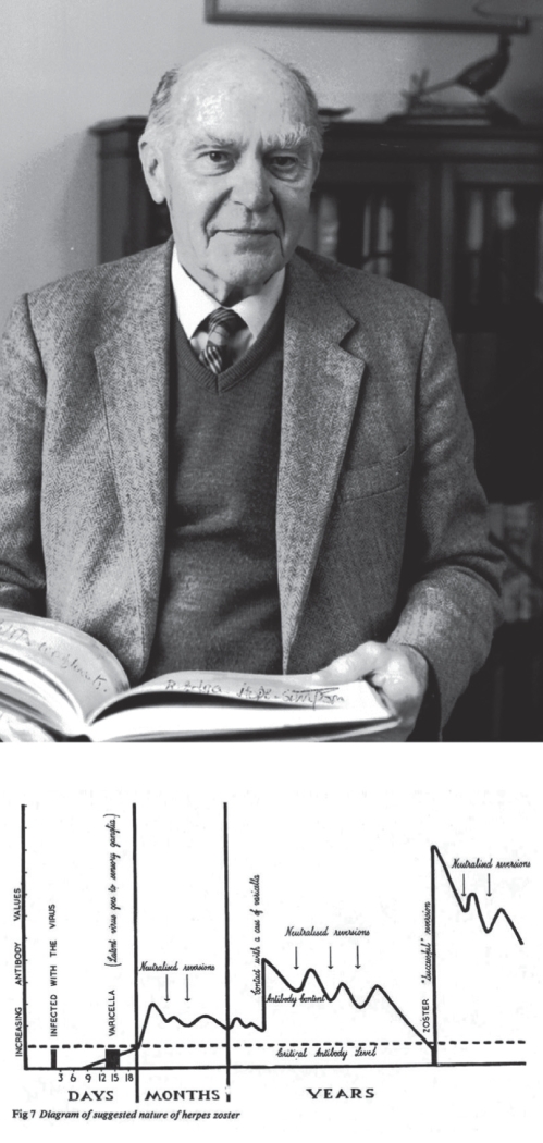
The history behind the current understanding of the varicella-zoster virus and its relationship to the pain conditions caused by shingles and postherpetic neuralgia are reviewed. The framework for the current conceptualization is Hope-Simpson's latency hypothesis. Data from recent work in virology, neuroanatomy and epidemiology are reviewed, as is work using varicella-zoster virus-infected animals. The recent data largely confirm Hope-Simpson's hypothesis and extend it significantly.

OBJECTIFS :: On passe en revue l’histoire qui a mené à la compréhension actuelle du virus varicelle-zona et de son lien avec les douleurs causées par l’algie post-zostérienne. L’hypothèse de latence de Hope-Simpson forme le cadre actuel de conceptualisation. On examine les données tirées de récents travaux en virologie, en neuroanatomie et en épidémiologie, de même que de travaux faisant appel à des animaux infectés par le virus varicelle-zona. Les données récentes confirment largement l’hypothèse de Hope-Simpson et la poussent beaucoup plus loin.


Figures

Figure 1)
Figure 1)
Top: R Edgar Hope-Simpson at the age of 90. Hope-Simpson’s epidemiological work was centred in Cirencester (United Kingdom), but his insights on the nature of varicella-zoster infection were the result of studying the isolated population of the Shetland island of Yell (United Kingdom). Bottom: The original schematic diagram of Hope-Simpson’s latency hypothesis (16)
Figure 2)
Figure 2)
Section of a punch biopsy from normal human skin stained with the pan-neuronal marker protein gene product 9.5. Nerve fibre bundles (green and yellow) course within the superficial dermis beneath the basement membrane (red) that separates the dermis from the epidermis (blue). Individual sensory axons (white) leave the dermal bundles, ascend, penetrate the basement membrane and ramify within the epidermis. Counting these intraepidermal nerve fibres affords a precise way to quantify the degree of sensory denervation following an attack of shingles. Reproduced from reference with permission
Figure 3)
Figure 3)
Left Panel: Plate 1 from Head and Campbell’s 1900 paper (8) showing the inflammatory and hemorrhagic effects of acute shingles. These drawings were made with a camera lucida drawing tube attached to the microscope and were subsequently hand-coloured. They are among the finest medical illustrations of the pre-Photoshop era. Top: Longitudinal section through the seventh thoracic dorsal root ganglia (DRG) from the affected side of a patient who died eight days after the onset of shingles. Extensive hemorrhage is seen in the dorsal part of the DRG that contains the cell bodies of primary afferent somatosensory neurons. Bottom: DRG sections from the seventh thoracic ganglia of a patient who died five months after the onset of shingles. The normal side is shown on the left and the affected side with extensive scarring is shown on the right. Shingles is rarely a fatal disease, but AW Campbell was a pathologist for the Rainhill County Asylum, which cared for the elderly poor and afforded ample material for study. Right panel: Section of the thoracic spinal cord (top) and eighth thoracic DRG (bottom) of a patient who died after a five-year history of severe postherpetic neuralgia. The spinal cord posterior horn is atrophied (arrows) on the affected side. Similar atrophy ran for seven spinal segments, although only one of the patient’s DRGs showed the characteristic scarring caused by shingles. Before this finding, extensive central nervous system pathology was not known to be present in postherpetic neuralgia. The lower right portion of the DRG contains normal neuronal cell bodies (white and red), but cells are missing throughout the fibrotic remainder. Reproduced from reference with permission

References

    1. Gilron I, Watson CPN, Cahill CM, Moulin DE. Neuropathic pain: A practical guide for the clinician. CMAJ. 2006;175:265–75. - PMC - PubMed
    1. Harpaz R, Ortega-Sanchez IR, Seward JF. Prevention of herpes zoster: Recommendations of the Advisory Committee on Immunization Practices (ACIP) MMWR Recomm Rep. 2008;57(RR-5):1–30. - PubMed
    1. Opstelten W, Eekhof J, Neven AK, Verheij T. Treatment of herpes zoster. Can Fam Physician. 2008;54:373–7. - PMC - PubMed
    1. Wu CL, Raja SN. An update on the treatment of postherpetic neuralgia. J Pain. 2008;9(Suppl 1):S19–30. - PubMed
    1. Jung BF, Johnson RW, Griffin DR, Dworkin RH. Risk factors for postherpetic neuralgia in patients with herpes zoster. Neurology. 2004;62:1545–51. - PubMed