. 2009 Sep;60(9):2633-43.
doi: 10.1002/art.24763.

HLA-B27 misfolding and the unfolded protein response augment interleukin-23 production and are associated with Th17 activation in transgenic rats

Affiliations

HLA-B27 misfolding and the unfolded protein response augment interleukin-23 production and are associated with Th17 activation in transgenic rats

Monica L DeLay et al. Arthritis Rheum. 2009 Sep.

Abstract

Objective: To determine whether HLA-B27 misfolding and the unfolded protein response (UPR) result in cytokine dysregulation and whether this is associated with Th1 and/or Th17 activation in HLA-B27/human beta(2)-microglobulin (Hubeta(2)m)-transgenic rats, an animal model of spondylarthritis.

Methods: Cytokine expression in lipopolysaccharide (LPS)-stimulated macrophages was analyzed in the presence and absence of a UPR induced by chemical agents or by HLA-B27 up-regulation. Cytokine expression in colon tissue and in cells purified from the lamina propria was determined by real-time reverse transcription-polymerase chain reaction analysis, and differences in Th1 and Th17 CD4+ T cell populations were quantified after intracellular cytokine staining.

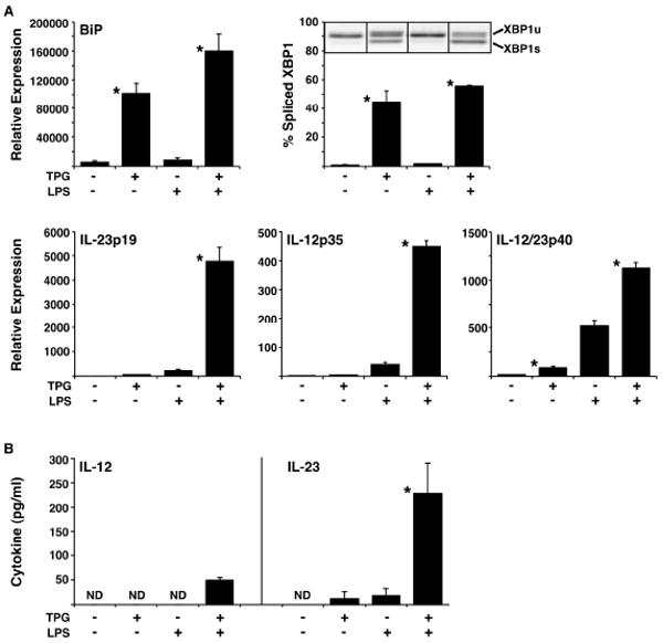
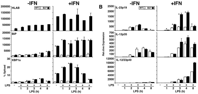
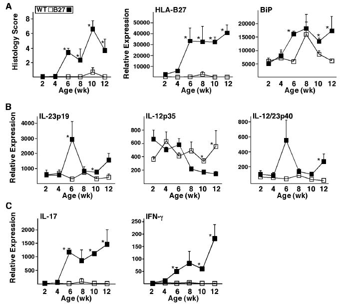
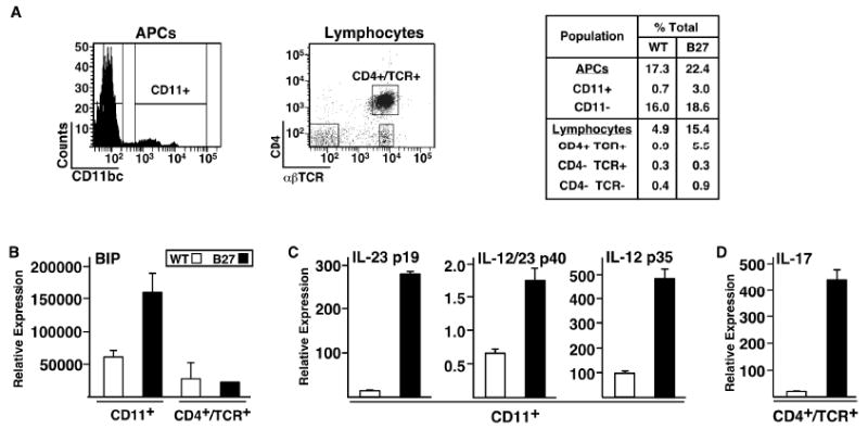
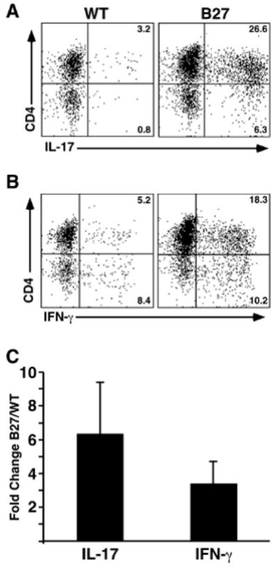
Results: Interleukin-23 (IL-23) was found to be synergistically up-regulated by LPS in macrophages undergoing a UPR induced by pharmacologic agents or by HLA-B27 misfolding. IL-23 was also increased in the colon tissue from B27/Hubeta(2)m-transgenic rats concurrently with the development of intestinal inflammation, and IL-17, a downstream target of IL-23, exhibited robust up-regulation in a similar temporal pattern. IL-23 and IL-17 transcripts were localized to CD11+ antigen-presenting cells and CD4+ T cells, respectively, from the colonic lamina propria. Colitis was associated with a 6-fold expansion of CD4+ IL-17-expressing T cells.

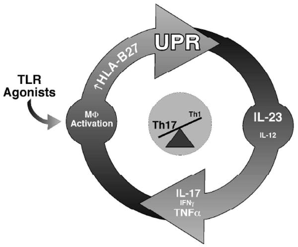
Conclusion: The IL-23/IL-17 axis is strongly activated in the colon of B27/Hubeta(2)m-transgenic rats with spondylarthritis-like disease. HLA-B27 misfolding and UPR activation in macrophages can result in enhanced induction of the pro-Th17 cytokine IL-23. These results suggest a possible link between HLA-B27 misfolding and immune dysregulation in this animal model, with implications for human disease.


Conflict of interest statement

Disclosures

The authors have no financial conflict of interest.

Figures

Figure 1
Figure 1
LPS-stimulated IL-23 production by macrophages is augmented by UPR activation. A, The UPR was induced in BM macrophages from WT rats by 1 μM TPG for 4 hours, with or without the addition of 10 ng/ml of LPS for the final 3 hours. Expression of BiP, IL-23p19, IL-12p35, and IL-12/23p40 mRNA was quantified by real time PCR normalized to β-actin. XBP1 splicing was determined by electrophoresis of RT-PCR products. Representative image of unspliced (upper band; XBP1u) and spliced (lower band; XBP1s) XBP1 PCR products are shown. Bars represent the mean of biological triplicates and are representative of at least three separate experiments. B, The UPR was induced in mouse BM macrophages by 1 μM TPG for 2 hours. This was followed by a 7 h washout period, after which cells were treated without or with 10 ng/ml of LPS for 24 hours. Supernatants were collected, and levels of IL-12 and IL-23 measured by ELISA. Bars represent the mean of triplicates and are representative of at least three separate experiments. Error bars represent standard error of the mean and statistical significance was determined using a standard t-test, where p <0.05 is considered significant as indicated by the asterisks (*).
Figure 2
Figure 2
LPS-induced IL-23p19 expression is augmented in the presence of a UPR in HLA-B27 transgenic rat macrophages. BM macrophages from WT and HLA-B27 transgenic (B27) rats were treated without or with 100 U/ml of recombinant rat IFN-γ for 20 h prior to stimulation with LPS for the number of hours shown. RNA expression for the indicated targets was quantified by real time PCR using β-actin for normalization, except for XBP1, which is shown as percent of spliced (XBP1s) XBP1 PCR products. A, Relative expression of HLA-B, BiP and percent spliced XBP1 mRNA. B, Relative expression of IL-23p19, IL-12p35 and IL-12/23p40 mRNA in BM macrophages. Bars indicate the mean of triplicate biological replicates and are representative of at least five separate experiments. Error bars represent standard error of the mean. The asterisk (*) indicates differences that are statistically significant (p < 0.05).
Figure 3
Figure 3
IL-23/IL-17 axis activation correlates with disease onset and progression in rat colon tissue. Histological assessment was performed and RNA was isolated from colon tissue of WT and HLA-B27 transgenic (B27) rats at 2, 4, 6, 8, 10 and 12 weeks of age. Transcript levels were analyzed by real time PCR and normalized to β-actin. A, Histological scores and relative expression of HLA-B27 and BiP in colon tissue. B, Relative expression of IL-23p19, IL-12p35 and IL-12/23p40 mRNA in colon tissue. C, Relative expression of IL-17 and IFN-γ mRNA in colon tissue. Data points represent the mean of measurements from 3 animals performed in duplicate. Error bars represent standard error of the mean. The asterisk (*) indicates differences that are statistically significant (p < 0.05).
Figure 4
Figure 4
Localization of IL-17 expression to CD4+/TCRαβ+ cells in HLA-B27 transgenic rat colonic lamina propria. Lamina propria cells were isolated from WT and HLA-B27 transgenic rat colon and further subdivided using FACS with antibodies against the indicated antigens. RNA was isolated from the sorted populations and analyzed by real time PCR to quantify transcript levels. A, Populations designated APCs and Lymphocytes were identified based on forward and side scatter characteristics. These populations were subdivided based on cell surface staining for the indicated antigens. B, C, and D, Transcript levels for the indicated genes comparing populations of either CD11+ cells or CD4+/TCR+ cells from WT and HLA-B27 transgenic rats. Data points represent the mean of 2-3 samples. Error bars represent standard error of the mean.
Figure 5
Figure 5
Expansion of CD4+ T cells expressing IL-17 in colonic lamina propria from HLA-B27 transgenic rats. Lamina propria lymphocytes were isolated from WT and HLA-B27 transgenic colon on a percoll gradient. Cells were analyzed for cell surface antigen and intracellular cytokines using FACS. Cells were gated on CD3+ and compared for cytokine production. A, Percentage of CD4+/IL-17+ and CD4-/IL-17+ cells in WT and HLA-B27 transgenic rats. B, Percentage of CD4+/IFN-γ+ and CD4-/IFN-γ+ cells in WT and HLA-B27 transgenic rats. C, Average fold-change in percentage of IL-17-expressing CD4+ T cells (IL-17; CD4+/IL-17+) and IFN-γ-expressing CD4+ T cells (IFN-γ; CD4+/IFN-γ+) populations between WT and HLA-B27 transgenic rats. Data shown are representative of three separate experiments using at least two animals per genotype and error bars represent standard error of the mean. Th17 cells are increased ∼6.3-fold (CI 3.1), and Th1 ∼3.4-fold (CI 1.3).
Figure 6
Figure 6
Proposed mechanism linking UPR activation as a consequence of HLA-B27 misfolding to activation of the IL-23/Il-17 axis. HLA-B27 upregulation may occur initially as a result of antigen presenting cell stimulation with TLR agonists from commensal microorganisms and/or low level IFN-γ production from innate immune cells such as NK cells. UPR activation is superimposed on macrophage activation because of HLA-B27 misfolding, resulting in greater IL-23 production in response to TLR agonists. This, in turn, stimulates Th17 cells to produce IL-17. Th1 activation, and/or double positive IL-17/IFN-γ producing cells, may help to sustain HLA-B27 expression thus perpetuating this cycle.

References

    1. Taurog JD, Maika SD, Satumtira N, Dorris ML, McLean IL, Yanagisawa H, et al. Inflammatory disease in HLA-B27 transgenic rats. Immunol Rev. 1999;169:209–223. - PubMed
    1. Smith JA, Marker-Hermann E, Colbert RA. Pathogenesis of ankylosing spondylitis: current concepts. Best Pract Res Clin Rheumatol. 2006;20(3):571–91. - PubMed
    1. May E, Dorris ML, Satumtira N, Iqbal I, Rehman MI, Lightfoot E, et al. CD8ab T cells are not essential to the pathogenesis of arthritis or colitis in HLA-B27 transgenic rats. J Immunol. 2003;170:1099–1105. - PubMed
    1. Dangoria NS, DeLay ML, Kingsbury DJ, Mear JP, Uchanska-Ziegler B, Ziegler A, et al. HLA-B27 misfolding is associated with aberrant intermolecular disulfide bond formation (dimerization) in the endoplasmic reticulum. J Biol Chem. 2002;277:23459–23468. - PubMed
    1. Antoniou AN, Ford S, Taurog JD, Butcher GW, Powis SJ. Formation of HLA-B27 homodimers and their relationship to assembly kinetics. J Biol Chem. 2004;279(10):8895–902. - PubMed

Publication types

MeSH terms