

Image-guided convection-enhanced delivery of muscimol to the primate brain
- PMID: 19715424
- PMCID: PMC2853729
- DOI: 10.3171/2009.7.JNS09652
Image-guided convection-enhanced delivery of muscimol to the primate brain
Abstract
Object: Muscimol is a potent gamma-aminobutyric acid-A receptor agonist that temporarily and selectively suppresses neurons. Targeted muscimol suppression of neuronal structures could provide insight into the pathophysiological processes and treatment of a variety of neurological disorders. To determine if muscimol delivered to the brain by convection-enhanced delivery could be monitored using a coinfused surrogate MR imaging tracer, the authors perfused the striata of primates with tritiated muscimol and Gd-diethylenetriamine pentaacetic acid (DTPA).
Methods: Three primates underwent convective coinfusion of (3)H-muscimol (0.8 microM) and Gd-DTPA (5 mM) into the bilateral striata. Primates underwent serial MR imaging during infusion, and the animals were killed immediately after infusion. Postmortem quantitative autoradiography and histological analysis was performed.
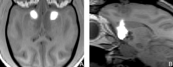
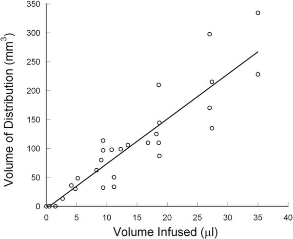
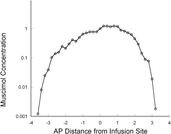
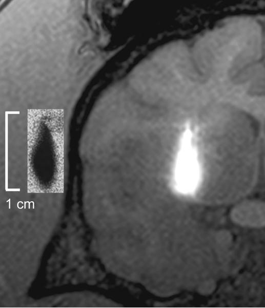
Results: Real-time MR imaging revealed that infusate (tritiated muscimol and Gd-DTPA) distribution was clearly discernible from the noninfused parenchyma. Real-time MR imaging of the infusion revealed the precise region of anatomical perfusion in each animal. Imaging analysis during infusion revealed that the distribution volume (Vd) of infusate linearly increased (R = 0.92) with volume of infusion (Vi). Overall, the mean (+/- SD) Vd/Vi ratio was 8.2 +/- 1.3. Autoradiographic analysis revealed that MR imaging of Gd-DTPA closely correlated with the distribution of (3)H-muscimol, and precisely estimated its Vd (mean difference in Vd, 7.4%). Quantitative autoradiograms revealed that muscimol was homogeneously distributed over the perfused region in a square-shaped concentration profile.
Conclusions: Muscimol can be effectively delivered to clinically relevant volumes of the primate brain. Moreover, the distribution of muscimol can be tracked using coinfusion of Gd-DTPA and MR imaging. The ability to perform accurate monitoring and to control the anatomical extent of muscimol distribution during its convection-enhanced delivery will enhance safety, permit correlations of muscimol distribution with clinical effect, and should lead to an improved understanding of the pathophysiological processes underlying a variety of neurological disorders.
Figures
References
-
- Baraldi M, Grandison L, Guidotti A. Distribution and metabolism of muscimol in the brain and other tissues of the rat. Neuropharmacology. 1979;18:57–62. - PubMed
-
- Cavagna FM, Maggioni F, Castelli PM, Dapra M, Imperatori LG, Lorusso V, et al. Gadolinium chelates with weak binding to serum proteins. A new class of high-efficiency, general purpose contrast agents for magnetic resonance imaging. Invest Radiol. 1997;32:780–796. - PubMed
-
- Chen MY, Lonser RR, Morrison PF, Governale LS, Oldfield EH. Variables affecting convection-enhanced delivery to the striatum: a systematic examination of rate of infusion, cannula size, infusate concentration, and tissue-cannula sealing time. J Neurosurg. 1999;90:315–320. - PubMed
Publication types
MeSH terms
Substances
Grants and funding
LinkOut - more resources
Full Text Sources
Other Literature Sources
Medical
