. 2009 Aug 28:7:56.
doi: 10.1186/1741-7007-7-56.

Golgi localisation of GMAP210 requires two distinct cis-membrane binding mechanisms

Affiliations

Golgi localisation of GMAP210 requires two distinct cis-membrane binding mechanisms

Jesus Cardenas et al. BMC Biol. .

Abstract

Background: The Golgi apparatus in mammals appears as a ribbon made up of interconnected stacks of flattened cisternae that is positioned close to the centrosome in a microtubule-dependent manner. How this organisation is achieved and retained is not well understood. GMAP210 is a long coiled-coil cis-Golgi associated protein that plays a role in maintaining Golgi ribbon integrity and position and contributes to the formation of the primary cilium. An amphipathic alpha-helix able to bind liposomes in vitro has been recently identified at the first 38 amino acids of the protein (amphipathic lipid-packing sensor motif), and an ARF1-binding domain (Grip-related Arf-binding domain) was found at the C-terminus. To which type of membranes these two GMAP210 regions bind in vivo and how this contributes to GMAP210 localisation and function remains to be investigated.

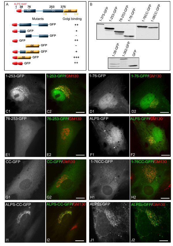
Results: By using truncated as well as chimeric mutants and videomicroscopy we found that both the N-terminus and the C-terminus of GMAP210 are targeted to the cis-Golgi in vivo. The ALPS motif was identified as the N-terminal binding motif and appeared concentrated in the periphery of Golgi elements and between Golgi stacks. On the contrary, the C-terminal domain appeared uniformly distributed in the cis-cisternae of the Golgi apparatus. Strikingly, the two ends of the protein also behave differently in response to the drug Brefeldin A. The N-terminal domain redistributed to the endoplasmic reticulum (ER) exit sites, as does the full-length protein, whereas the C-terminal domain rapidly dissociated from the Golgi apparatus to the cytosol. Mutants comprising the full-length protein but lacking one of the terminal motifs also associated with the cis-Golgi with distribution patterns similar to those of the corresponding terminal end whereas a mutant consisting in fused N- and C-terminal ends exhibits identical localisation as the endogenous protein.

Conclusion: We conclude that the Golgi localisation of GMAP210 is the result of the combined action of the two N- and C-terminal domains that recognise different sub-regions of the cis-GA. Based on present and previous data, we propose a model in which GMAP210 would participate in homotypic fusion of cis-cisternae by anchoring the surface of cisternae via its C-terminus and projecting its distal N-terminus to bind the rims or to stabilise tubular structures connecting neighbouring cis-cisternae.


Figures

Figure 1
Figure 1
The N-terminal domain of GMAP210 targets early compartments of the secretory pathway. (A) Confocal images of RPE1 cells transfected with the Nter-GFP construct (A1) and double labelled for GM130 (A2, merged image) and Golgin245 (A3, merged image). An enlarged view of A1 is shown at the bottom. Insets show enlarged views. In bottom panels, fluorescence intensity profiles of lines drawn in insets are shown. (B) Nter-GFP transfected RPE1 cells treated with nocodazole and stained for GM130 and Golgin245. Bottom panels show an enlarged view (left) and fluorescence intensity profiles (right) of the line drawn over a nocodazole-induced Golgi element. (C) Dynamics of the NterDsRed fusion protein in a RPE1 cell line stably expressing GT™-GFP. Selected frames at the indicated times from Movie 1 (in Additional file 1) are shown. An enlarged view is shown at the bottom. Arrows point to sites in which the NterDsRed protein is located between two membrane elements. (D) RPE1 cells treated with siRNA against GMAP210 for 72 h were transfected with the Nter-GFP construction (D1), incubated for 16 h and then fixed and immuno-stained for GM130 in red and GMAP210 in blue (D2). ND-NT indicates a non-depleted-non-transfected cell, D-NT a depleted non-transfected cell, and D-T a depleted and transfected cell. (E, F) Nter-GFP transfected cells (E) were treated with BFA (F) and then stained for Sec31 to reveal ER exit sites. Enlarged views of individual labellings are shown at right. Arrows indicate ERES containing the GMAP210 N-terminal domain. Bars = 5 μm
Figure 2
Figure 2
The ALPS motif of GMAP210 is sufficient for Golgi binding. (A, B) Schematic representation of the GFP-tagged N-terminal domain of GMAP210 and all the truncated or fusion constructs in this domain that were tested for their binding to GA (A) and their migration on SDS-PAGE as revealed by Western blot with an anti-GFP antibody after expression in RPE1 cells (B). The ALPS-like motif is represented in red and coiled-coil regions in light blue. The yellow rectangle corresponds to the continuous coiled-coil domain used in the indicated constructs. (C to J) Subcellular localisation of the different N-terminal mutants and other constructs (as indicated) expressed in RPE1 cells and double labelled for GM130. Arrows in (F1) indicate ERES. Bars = 10 μm.
Figure 3
Figure 3
A detailed analysis of the distribution of the ALPS-CC mutant protein in Golgi membranes. (A, B) Cells transfected with the ALPS-CC-GFP construct (A) were treated (B) with nocodazole and labelled with anti-GM130 and anti-Golgin245 antibodies. Merged images are shown. Enlarged views of insets show merged images of GFP and Golgin245 labellings in left panels, merged images of GM130 and Golgin245 labellings in middle panels and merged images of the three labellings in right panels. At the bottom, fluorescence intensity profiles of lines drawn over Golgi membranes revealed cis-Golgi localisation of the mutant (lane 1) but also a striking enrichment in regions from which GM130 is excluded and vice versa (arrows and lanes 2 and 3). Bars = 5 μm.
Figure 4
Figure 4
The C-terminal region of GMAP210 targets the cis-Golgi compartment and the centrosome. (A, B) RPE1 cells were transfected with the C-terminal domain of GMAP210 (aa 1778–1979) in fusion with GFP, fixed and double labelled for GM130 and endogenous GMAP210 (A) or for the centrosomal protein ninein and GMAP210 (B). Merged images are shown in A2 and B2. Arrows indicate the centrosome. (C) A merged image of GMAP210-depleted cells transfected with the GFP-Cter construct and stained for GM130 and endogenous GMAP210. ND-NT indicates a non-depleted non-transfected cell, D-NT a depleted non-transfected cell and D-T, a depleted and transfected cell. (D to F) Confocal images showing the distribution of GFP-Cter fusion protein with respect to the cis-Golgi marker GM130 (D), the medial Golgi marker CTR433 (E) or the trans-Golgi marker Golgin 245 (F). Corresponding fluorescence intensity profiles are shown at right. (G) A GFP-Cter transfected cell treated with nocodazole and labelled for GM130. Enlarged views of single labellings are shown at the bottom. (H) Live imaging of a GFP-Cter transfected cell treated with BFA. BFA was added to a final concentration of 2.5 μg/ml at time 5.0 seconds and images were taken with 0.2-second intervals. Selected frames of Movie 2 (in Additional file 2) at indicated time points are shown. Note that GFP-Cter rapidly dissociated from membranes after BFA addition whereas fluorescence at the centrosome decreased only slightly (arrow). Bars = 5 μm.
Figure 5
Figure 5
The N-terminus and the C-terminus of GMAP210 bind different regions of the Golgi ribbon. (A to C) Nter-DsRed (1–375 aas) and GFP-Cter (1778–1979 aas) constructs were co-transfected in RPE1 cells and their distribution analysed by confocal microscopy (A) or deconvolution (B, C). Merged images of the two constructs (A) or of the two constructs with giantin (B) or with endogenous GMAP210 (C) immunolocalisation are shown. High magnifications of the selected regions in (A) and (B) show individual labellings and the merge. Arrows indicate identical locations for each panel. (D) RPE1 cells co-transfected with Nter-DsRed and GFP-Cter and treated with BFA. All GFP-Cter becomes cytosolic except at the centrosome (arrow). (E, F) Time-lapse analysis of RPE1 cells transiently transfected with Nter-DsRed and GFP-Cter in control conditions or after nocodazole treatment. Selected frames of Movies 3 (control) or 4 (nocodazole) (see Additional files 3 and 4, respectively) are shown at the same time points. At right, enlarged views of selected areas are shown. (G) RPE1 cells transfected with a truncated mutant consisting of both N- and C-termini expressed in fusion (Nter-Cter-GFP, G1) and double labelled for endogenous GMAP210 (G2). A merged image is shown in G3. In (H), a Nter-Cter-GFP transfected cell was treated with nocodazole and triple labelled for endogenous GMAP210 and Golgin245. High magnifications of the selected region at right show individual labellings and the merge. Arrows indicate identical localisation of the truncated mutant and the endogenous protein in isolated Golgi ministacks. Bars = 5 μm.
Figure 6
Figure 6
Contribution of both Golgi binding domains to the localisation of GMAP210. (A) Schematic representation of the full-length GMAP210 protein and of the truncated mutants used in this experiment. The ALPS-like motif is represented in red, coiled-coil regions in light blue, and the predicted GRAB domain in green. Western blot of all of these constructs expressed in RPE1 cells is presented in (F). (B) Cells expressing the mutant lacking the N-terminal domain (GFP-GMAPΔN (251–1979 aas)) were stained for GM130 (B2) and Golgin245 (B3). Insets show enlarged views. Fluorescence intensity profiles at the bottom revealed perfect co-localisation of this mutant with the cis-Golgi marker GM130. (C) Cells transfected with the mutant lacking the C-terminal domain (GFP-GMAPΔC (1–1778 aas)) were processed as above. Although the mutant mostly associated with the cis-side of the GA, it was also present in areas that did not contain GM130. In addition, it also accumulates in some tubular and vesicular structures devoid of GM130. (D, E) The central coiled-coil domains do not contain Golgi targeting information. Cells expressing the GFP-GMAPCC1 (D) or GFP-GMAPCC2 fusion proteins and labelled for GM130 are shown. Bars = 5 μm.

References

    1. Polishchuk RS, Mironov AA. Structural aspects of Golgi function. Cell Mol Life Sci. 2004;61:146–158. doi: 10.1007/s00018-003-3353-8. - DOI - PMC - PubMed
    1. Rios RM, Bornens M. The Golgi apparatus at the cell centre. Curr Opin Cell Biol. 2003;15:60–66. doi: 10.1016/S0955-0674(02)00013-3. - DOI - PubMed
    1. Caviston JP, Holzbaur EL. Microtubule motors at the intersection of trafficking and transport. Trends Cell Biol. 2006;16:530–537. doi: 10.1016/j.tcb.2006.08.002. - DOI - PubMed
    1. Thyberg J, Moskalewski S. Role of microtubules in the organization of the Golgi complex. Exp Cell Res. 1999;246:263–279. doi: 10.1006/excr.1998.4326. - DOI - PubMed
    1. Burkhardt JK, Echeverri CJ, Nilsson T, Vallee RB. Overexpression of the dynamitin (p50) subunit of the dynactin complex disrupts dynein-dependent maintenance of membrane organelle distribution. J Cell Biol. 1997;139:469–484. doi: 10.1083/jcb.139.2.469. - DOI - PMC - PubMed

Publication types

MeSH terms