. 2009 Aug 28:6:11.
doi: 10.1186/1742-4933-6-11.

Effect of ageing on CMV-specific CD8 T cells from CMV seropositive healthy donors

Affiliations

Effect of ageing on CMV-specific CD8 T cells from CMV seropositive healthy donors

María Luisa Pita-Lopez et al. Immun Ageing. .

Abstract

Background: Ageing is associated with changes in the immune system with substantial alterations in T-lymphocyte subsets. Cytomegalovirus (CMV) is one of the factors that affect functionality of T cells and the differentiation and large expansions of CMV pp65-specific T cells have been associated with impaired responses to other immune challenges. Moreover, the presence of clonal expansions of CMV-specific T cells may shrink the available repertoire for other antigens and contribute to the increased incidence of infectious diseases in the elderly. In this study, we analyse the effect of ageing on the phenotype and frequency of CMV pp65-specific CD8 T cell subsets according to the expression of CCR7, CD45RA, CD27, CD28, CD244 and CD85j.

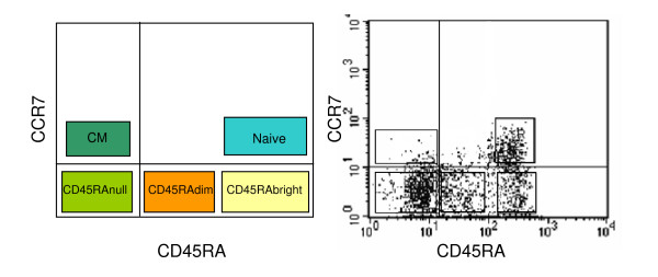
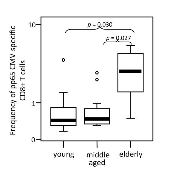
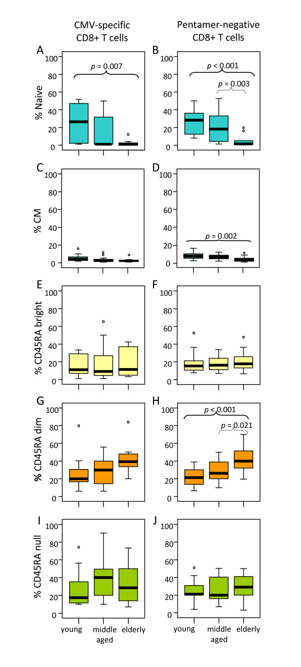
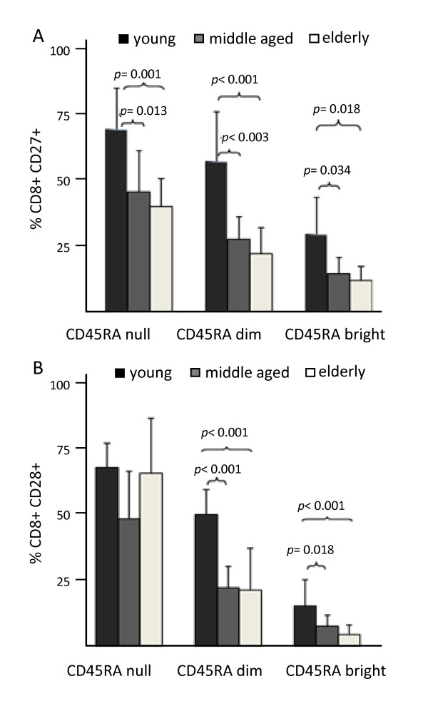
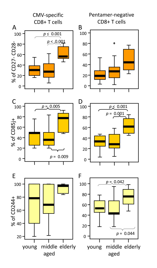
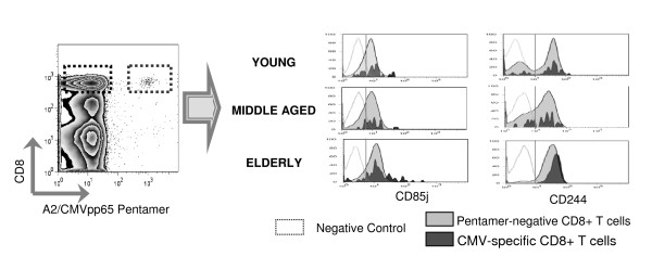
Results: Peripheral blood from HLA-A2 healthy young, middle-aged and elderly donors was analysed by multiparametric flow cytometry using the HLA-A*0201/CMV pp65(495-504) (NLVPMVATV) pentamer and mAbs specific for the molecules analysed. The frequency of CMV pp65-specific CD8 T cells was increased in the elderly compared with young and middle-aged donors. The proportion of naïve cells was reduced in the elderly, whereas an age-associated increase of the CCR7(null) effector-memory subset, in particular those with a CD45RA(dim) phenotype, was observed, both in the pentamer-positive and pentamer-negative CD8 T cells. The results also showed that most CMV pp65-specific CD8 T cells in elderly individuals were CD27/CD28 negative and expressed CD85j and CD244.

Conclusion: The finding that the phenotype of CMV pp65-specific CD8 T cells in elderly individuals is similar to the predominant phenotype of CD8 T cells as a whole, suggests that CMV persistent infections contributes to the age-related changes observed in the CD8 T cell compartment, and that chronic stimulation by other persistent antigens also play a role in T cell immunosenescence. Differences in subset distribution in elderly individuals showing a decrease in naive and an increase in effector-memory CD8 T cells may be relevant in the age-associated defective immune response.


Figures

Figure 1
Figure 1
CD8 T cell-subset distribution according to the expression of CCR7 and CD45RA. Schematic representation (left) and a representative experiment (right) of the five CD8 T cell subsets that can be defined by the analysis of CCR7 and CD45RA markers.
Figure 2
Figure 2
Elderly individuals show an increased frequency of CMVpp65-specific CD8 T cells. The frequency of A2/CMVpp65 pentamer-positive CD8 T cells was analysed in young, middle aged and elderly donors. Data are presented as diagrams of boxes; the lower boundary of the box indicates the 25th percentile and the upper boundary the 75th percentile. Error bars above and below the box indicate the 90th and 10th percentiles. A line within the box marks the median. Outliers are represented as individual points. P values lower than 0.05 were considered statistically significant.
Figure 3
Figure 3
Influence of age on CD8 T cell subset distribution. According to the subset model depicted in Figure 1, CD8 T cells were subdivided into naive, CM, EMRAbright, EMRAdim, EMRAnull cells and analysed in young, middle aged and elderly individuals. Results for CMV pentamer-positive (left) and pentamer-negative (right) CD8 T cells are shown. Data are presented as diagrams of boxes as indicated in Figure 2 legend.
Figure 4
Figure 4
Expression of CD27 and CD28 on the effector-memory CD8 T cell subsets. EM CD8 T cells defined as CCR7null were subdivided into further subsets according to the model in Figure 1 and analysed in young, middle aged and elderly individuals. Columns and bars represent the mean and SD.
Figure 5
Figure 5
The majority of A2/CMV-pp65 pentamer-positive CD8 T cells from elderly individuals display a CD27- CD28- phenotype and express CD85j and CD244. The percentage of CD27-CD28- (A), CD85j+ (B) and CD244+ (C) CD8 T cells was analysed on PBMC gated on CMV pentamer-positive (left) and pentamer-negative (right) CD8 T cells in young, middle aged and elderly donors. Data are presented as diagrams of boxes as indicated in Figure 2 legend.
Figure 6
Figure 6
Analysis of CD85j and CD244 expression on CD8 T cells. Dot-plot chart illustrates how pentamer-positive and pentamer-negative CD8 T cells were selected. Histograms show the expression of CD85j and CD244 in A2/CMVpp65 pentamer-positive (dark grey) and pentamer-negative (light grey) CD8 T cells from representative young, middle-aged and elderly individuals. The empty histograms represent the negative isotype control for each experiment.

References

    1. Motta VN, Martins SL. Impairment of cytomegalovirus-specific cellular immune response as a risk factor for cytomegalovirus disease in transplant recipients. Braz J Med Biol Res. 2008;41:5–11. doi: 10.1590/S0100-879X2006005000193. - DOI - PubMed
    1. Allart S, Lule J, Serres B, Jones T, Davignon JL, Malecaze F, Davrinche C. Impaired killing of HCMV-infected retinal pigment epithelial cells by anti-pp65 CD8(+) cytotoxic T cells. Invest Ophthalmol Vis Sci. 2003;44:665–671. doi: 10.1167/iovs.02-0547. - DOI - PubMed
    1. Lang KS, Moris A, Gouttefangeas C, Walter S, Teichgraber V, Miller M, Wernet D, Hamprecht K, Rammensee HG, Stevanovic S. High frequency of human cytomegalovirus (HCMV)-specific CD8+ T cells detected in a healthy CMV-seropositive donor. Cell Mol Life Sci. 2002;59:1076–1080. doi: 10.1007/s00018-002-8488-5. - DOI - PMC - PubMed
    1. Chidrawar S, Khan N, Wei W, McLarnon A, Smith N, Nayak L, Moss P. Cytomegalovirus-seropositivity has a profound influence on the magnitude of major lymphoid subsets within healthy individuals. Clin Exp Immunol. 2009;155:423–432. doi: 10.1111/j.1365-2249.2008.03785.x. - DOI - PMC - PubMed
    1. Derhovanessian E, Larbi A, Pawelec G. Biomarkers of human immunosenescence: impact of Cytomegalovirus infection. Curr Opin Immunol. 2009;21:440–445. doi: 10.1016/j.coi.2009.05.012. - DOI - PubMed