. 2009 Aug 28:8:68.
doi: 10.1186/1476-4598-8-68.

Docetaxel-induced prostate cancer cell death involves concomitant activation of caspase and lysosomal pathways and is attenuated by LEDGF/p75

Affiliations

Docetaxel-induced prostate cancer cell death involves concomitant activation of caspase and lysosomal pathways and is attenuated by LEDGF/p75

Melanie Mediavilla-Varela et al. Mol Cancer. .

Abstract

Background: Hormone-refractory prostate cancer (HRPC) is characterized by poor response to chemotherapy and high mortality, particularly among African American men when compared to other racial/ethnic groups. It is generally accepted that docetaxel, the standard of care for chemotherapy of HRPC, primarily exerts tumor cell death by inducing mitotic catastrophe and caspase-dependent apoptosis following inhibition of microtubule depolymerization. However, there is a gap in our knowledge of mechanistic events underlying docetaxel-induced caspase-independent cell death, and the genes that antagonize this process. This knowledge is important for circumventing HRPC chemoresistance and reducing disparities in prostate cancer mortality.

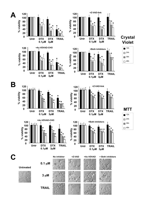
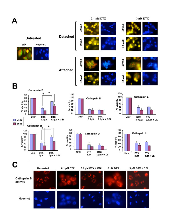
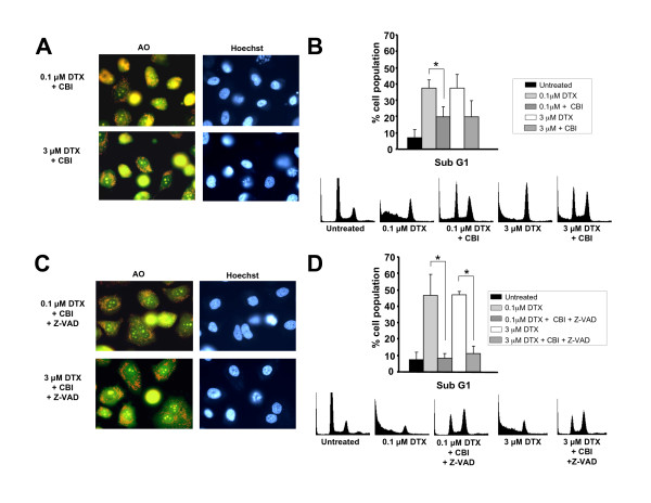
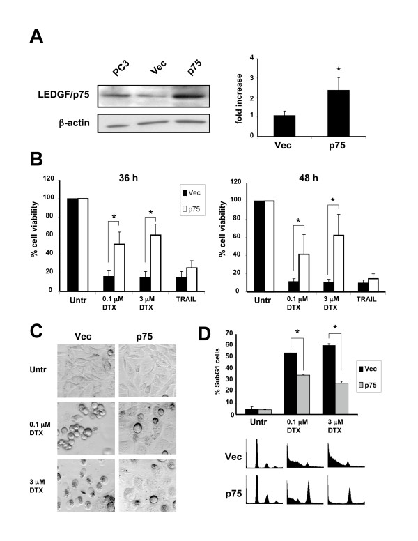
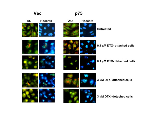
Results: We investigated mechanistic events associated with docetaxel-induced death in HRPC cell lines using various approaches that distinguish caspase-dependent from caspase-independent cell death. Docetaxel induced both mitotic catastrophe and caspase-dependent apoptosis at various concentrations. However, caspase activity was not essential for docetaxel-induced cytotoxicity since cell death associated with lysosomal membrane permeabilization still occurred in the presence of caspase inhibitors. Partial inhibition of docetaxel-induced cytotoxicity was observed after inhibition of cathepsin B, but not inhibition of cathepsins D and L, suggesting that docetaxel induces caspase-independent, lysosomal cell death. Simultaneous inhibition of caspases and cathepsin B dramatically reduced docetaxel-induced cell death. Ectopic expression of lens epithelium-derived growth factor p75 (LEDGF/p75), a stress survival autoantigen and transcription co-activator, attenuated docetaxel-induced lysosomal destabilization and cell death. Interestingly, LEDGF/p75 overexpression did not protect cells against DTX-induced mitotic catastrophe, and against apoptosis induced by tumor necrosis factor related apoptosis inducing ligand (TRAIL), suggesting selectivity in its pro-survival activity.

Conclusion: These results underscore the ability of docetaxel to induce concomitantly caspase-dependent and independent death pathways in prostate cancer cells. The results also point to LEDGF/p75 as a potential contributor to cellular resistance to docetaxel-induced lysosomal destabilization and cell death, and an attractive candidate for molecular targeting in HRPC.


Figures

Figure 1
Figure 1
Docetaxel (DTX) induces caspase-dependent death in PC3 cells. A. Caspase-3/7 and -2 activity assays in PC3 cells treated with DTX, TRAIL, or STS in the presence and absence of the pan caspase inhibitor Z-VAD-FMK and the caspase-2 inhibitor Ac-VDVAD-CHO respectively. Cells were pre-treated 1 h prior to drug treatment. Activity was determined by measuring the cleavage of the fluorogenic substrate Ac-DEVD-AMC for caspase-3/7 and Ac-VDVAD-AMC for caspase-2. Fold activation was determined by normalization of the test sample to untreated controls: B. Immunoblotting analysis of PARP and LEDGF/p75 cleavage in DTX-treated PC3 cells in the presence and absence of Z-VAD-FMK: C. Flow cytometric analysis of mitochondrial membrane potential (MMP), using the JC-1 method, in PC3 cells treated with DTX in the presence and absence of Z-VAD-FMK for 48 h. A representative of three independent experiments is shown: D. Cell cycle analysis of PC3 cells treated with DTX in the presence and absence of Z-VAD-FMK for 48 h. Flow cytometric analysis of cells stained with propidium iodide was used to determine the percentage of cells in SubG1 and G2/M. Representative DNA histograms are shown. Error bars in panels A and D represent the standard deviation of at least three independent experiments done in triplicates (* p < 0.05, t-test).
Figure 2
Figure 2
Inhibition of caspases does not block DTX-induced cytotoxicity in PC3 cells. A. Percentage of surviving PC3 cells treated with DTX in the presence and absence of Z-VAD-FMK, AC-VDVAD-CHO, or both inhibitors. Cells were pre-treated with the inhibitors 1 h before exposure to DTX. Cell viability was determined by crystal violet staining. Absorbance was measured at 570 nm and the values were normalized against those of untreated cells, which were assumed to be 100% viable. Errors bars represent the standard deviation of at least three independent experiments done in triplicate (*p < 0.05 compared to untreated, t-test): B. Same as in panel A except that cell viability was measured using the MTT assay. Errors bars represent the standard deviation of two independent experiments done in hexuplicates (*p < 0.05 compared to untreated, t-test): C. Morphological analysis of PC3 cells treated as indicated above for 48 h. Cells were visualized on an Olympus IX70 inverted microscope equipped with Hoffmann Modulation Contrast.
Figure 3
Figure 3
DTX induces lysosomal membrane permeabilization (LMP) in PC3 cells. A. Determination of lysosomal integrity in PC3 cells treated with DTX for 24 hours in the presence and absence of Z-VAD-FMK. Cells were exposed to acridine orange (AO) for lysosome visualization under fluorescence microscopy. Untreated cells showed localized granular red fluorescence corresponding to staining of intact lysosomes. Cells that detached after treatment with DTX in the presence and absence of Z-VAD-FMK displayed increased yellow fluorescence indicative of LMP. Most cells that remained attached after treatment appeared to have intact lysosomes but exhibited multinucleation. Hoescht staining was used to visualize nuclear morphology: B. Cathepsin B inhibitor (CBI) but not inhibitors of cathepsin L (CLI) or cathepsin D (CDI) attenuated cell death induced by 0.1 μM DTX (top set of graphs) and 3 μM DTX (bottom set of graphs), as assessed by crystal violet viability assays. Errors bars represent the standard deviation of at least three independent experiments done in triplicate (*p < 0.05, t-test): C. Detection of intracellular cathepsin B activity in PC3 cells using the fluorogenic substrate Magic Red MR-(RR)2. Cells treated with DTX for 36 hours showed diffuse cathepsin B activity (red fluorescence), whereas the activity in untreated cells and cells treated with DTX in the presence of CBI was localized mainly to lysosomes.
Figure 4
Figure 4
Simultaneous inhibition of caspases and cathepsin B attenuates DTX-induced cell LMP and DNA fragmentation. A. PC3 cell cultures treated with DTX for 36 hours in the presence of cathepsin B inhibitor and stained with acridine orange (AO) appeared to have less detached yellow cells than cultures treated with DTX in the absence of inhibitor (compare with Fig. 3A). Images were acquired from fields that had both attached and detached cells: B. Cell cycle analysis of PC3 cells treated with DTX in the presence and absence of cathepisn B inhibitor for 48 h. Flow cytometric analysis of cells stained with propidium iodide was used to determine the percentage of subG1 cells. Error bars represent the standard deviation of at least three independent experiments (* p < 0.05, t-test). Representative DNA histograms for one of the experiments are shown: C. PC3 cell cultures treated with DTX for 36 hours in the presence of both Z-VAD-FMK and cathepsin B inhibitor, and stained with acridine orange, showed very few yellow detached cells, as compared with cultures treated with DTX in the absence of inhibitors (see Fig. 3A): D. Cell cycle analysis of PC3 cells treated with DTX in the presence and absence of Z-VAD-FMK and cathepisn B inhibitor for 48 h. Flow cytometric analysis of cells stained with propidium iodide was used to determine the percentage of subG1 cells. Error bars represent the standard deviation of at least three independent experiments (* p < 0.05, t-test). Representative DNA histograms for one of the experiments are shown.
Figure 5
Figure 5
Overexpression of LEDGF/p75 attenuates DTX-induced cell death. A. LEDGF/p75 expression in parental PC3 cells, pools of PC3 clones stably transfected with empty pcDNA vector (Vec), or pcDNA-ledgfp75 (p75), analyzed by immunoblotting (right panel) and RT-PCR: B. Stable overexpression of LEDGF/p75 attenuated DTX-induced cytotoxicity after 36 and 48 h of exposure to low and high concentrations of the drug. Cell survival was determined by crystal violet staining. Data is representative of three independent experiments. Error bars represent the standard deviation of at least three independent experiments done in triplicates (* p < 0.05, t-test): C. Morphology of PC3 clones (Vec and p75) treated with DTX for 48 hours. Clones overexpressing LEDGF/p75 showed less dead and detached cells than clones transfected with empty vector: D. Cell cycle analysis of PC3 clones (Vec, and p75) treated with DTX for 48 h. Flow cytometric analysis of cells stained with propidium iodide was used to determine the percentage of subG1 cells. Overexpression of LEDGF/p75 significantly attenuated DTX-induced DNA fragmentation. Representative DNA histograms are shown. Error bars represent the standard deviation of at least three independent experiments done in triplicates (* p < 0.05, t-test).
Figure 6
Figure 6
Overexpression of LEDGF/p75 attenuates DTX-induced LMP. PC3 cell clones (Vec and p75) were treated with DTX for up to 48 hours and stained with acridine orange (AO) to visualize LMP. Clones stably transfected with empty vector (Vec) showed increased LMP as indicated by minimal or absence of punctuated lysosomal red fluorescence, associated with increased number of detached yellow cells. Clones overexpressing LEDGF/p75 showed less cell detachment and increased lysosomal stability after DTX treatment. These resistant clones displayed extensive multinucleation.

References

    1. Jemal A, Siegel R, Ward E, Hao Y, Xu J, Thun MJ. Cancer statistics, 2009. CA Cancer J Clin. 2009;59:225–49. doi: 10.3322/caac.20006. - DOI - PubMed
    1. Peters N, Armstrong K. Racial differences in prostate cancer treatment outcomes: a systematic review. Cancer Nursing. 2005;28:108–18. doi: 10.1097/00002820-200503000-00004. - DOI - PubMed
    1. Latini DM, Elkin EP, Cooperberg MR, Sadetsky N, Duchane J, Carroll PR. Differences in clinical characteristics and disease-free survival for Latino, African American, and non-Latino white men with localized prostate cancer: data from CaPSURE. Cancer. 2006;106:789–95. doi: 10.1002/cncr.21675. - DOI - PubMed
    1. Dagher R, Li N, Abraham S, Rahman A, Sridhara R, Pazdur R. Approval summary: Docetaxel in combination with prednisone for the treatment of androgen-independent hormone-refractory prostate cancer. Clin Cancer Res. 2004;10:8147–51. doi: 10.1158/1078-0432.CCR-04-1402. - DOI - PubMed
    1. So A, Hadaschik B, Sowery R, Gleave M. The role of stress proteins in prostate cancer. Curr Genomics. 2007;8:252–61. doi: 10.2174/138920207781386951. - DOI - PMC - PubMed

Publication types

MeSH terms