. 2009 Aug 28;35(4):403-13.
doi: 10.1016/j.molcel.2009.06.025.

TERRA RNA binding to TRF2 facilitates heterochromatin formation and ORC recruitment at telomeres

Affiliations

TERRA RNA binding to TRF2 facilitates heterochromatin formation and ORC recruitment at telomeres

Zhong Deng et al. Mol Cell. .

Abstract

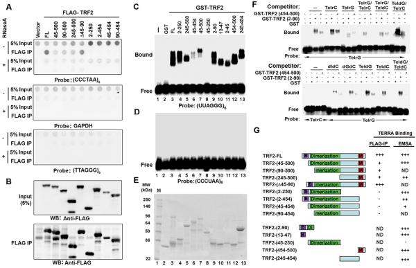
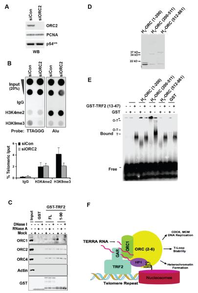
Telomere-repeat-encoding RNA (referred to as TERRA) has been identified as a potential component of yeast and mammalian telomeres. We show here that TERRA RNA interacts with several telomere-associated proteins, including telomere repeat factors 1 (TRF1) and 2 (TRF2), subunits of the origin recognition complex (ORC), heterochromatin protein 1 (HP1), histone H3 trimethyl K9 (H3 K9me3), and members of the DNA-damage-sensing pathway. siRNA depletion of TERRA caused an increase in telomere dysfunction-induced foci, aberrations in metaphase telomeres, and a loss of histone H3 K9me3 and ORC at telomere repeat DNA. Previous studies found that TRF2 amino-terminal GAR domain recruited ORC to telomeres. We now show that TERRA RNA can interact directly with the TRF2 GAR and ORC1 to form a stable ternary complex. We conclude that TERRA facilitates TRF2 interaction with ORC and plays a central role in telomere structural maintenance and heterochromatin formation.


Figures

Figure 1
Figure 1. Identification of TERRA RNA binding proteins using RNA affinity purification
A) Biotinylated RNA oligonucleotides (CACUGA)8, (UUAGGG)8, or (CCCUAA)8 were incubated with Raji cell nuclear extracts and then assayed for protein binding by silver staining of SDS-PAGE. LC/MS/MS identified polypeptides are indicated. B) Polypeptides enriched by (UUAGGG)8 RNA affinity were subject to identification by LC/MS/MS and validated by Western blot with antibodies to TRF2, hRap1, TRF1, ORC1, ORC2, ORC4, TIN2, PCNA, MCM5, RPA70, RPA34, or EBNA1, as indicated. Input nuclear extract (5%) is indicated at the left of each panel. C) Same as in B, except Western blot antibodies included MeCP2, DNA PKcs, PARP1, TopoI, Topo II, Blm, TRF2, Dyskerin, or Actin as indicated.
Figure 2
Figure 2. TRF1 and TRF2 bind TERRA RNA
A) RNA IP assays were performed in HCT116 cells transfected with FLAG-tagged TRF1, TRF2, hRap1, POT1, TPP1, or expression vector alone. Input (5%) is shown in the top two rows. IPs are shown in duplicates. IP recovered RNA was detected with probes containing the telomere C-rich strand (top panel), G-rich strand (middle panel), or GAPDH (lower panel). B) Western blot was used to detect the FLAG-tagged proteins used in experiments shown in panel A. Input and FLAG-IP material were detected by Western blot with antibodies to FLAG or loading control Actin. C) RNA IP assays were performed in HCT116 cells transfected with FLAG-tagged TRF1, TRF2, or expression vector alone. Bead-bound IP complex was subject to mock (−) or DNase I (+, 20U/100 μl) treatment for 30 mins at 37 °C. RNA was recovered after treatment, dot blotted and then visualized by hybridization with probes to CCCTAA, TTAGGG, or GAPDH sequence as indicated. D) RNA-ChIP assays were performed using HCT116 cells and rabbit polyclonal antibodies to TRF1, TRF2, hRap1, or control IgG. ChIP RNA was either mock treated (−) or treated with RNase A (200 μg/ml) or RNase H (30U/ml) prior to dot blot analysis. RNA was visualized by hybridization with probe containing the C-rich telomere repeat (top panel), GAPDH (middle panel) or the G-rich telomere repeat (lower panel). E) Same as in D, except using U2OS cells and an additional hybridization with a probe specific for U6 RNA. F) Quantification of RNA ChIP assays for each cell type represented by dot blots shown in panels D and E. Values are the means and SD (error bars) for at least three independent experiments.
Figure 3
Figure 3. Multiple domains in TRF2 bind TERRA RNA
A) RNA IP assay was performed in HCT116 cells transfected with FLAG-tagged TRF2 full length (FL), aa45-500, 90-500, 245-500, Δ45-90, 2-250, 2-454, 45-454, 90-454 or vector control. RNA isolated from FLAG-IP was detected by dot blot hybridization with probe containing the C-rich telomere repeat (top panel), G-rich telomere repeat (middle panel), or GAPDH (lower panel). FLAG-IP RNA was either mock (−) treated or treated with RNase A (+) prior to dot blotting. Top row of each sample represent 5% of the input RNA. B) FLAG-TRF2 protein expression levels (top panel) and immunoprecipitation efficiency (lower panel) were assayed by Western blot as control for experiments shown in panel A. C) Agarose EMSA was used to measure TRF2 RNA binding activity in vitro. GST, GST-TRF2 (full length), GST-TRF2 (2-250), (245-500), (45-454), (45-500), (45-250), (2-90), (13-47), (2-45), (454-500), or (245-454) were assayed for binding to 32P-labeled RNA oligonucleotides for (UUAGGG)6. D) Same as in C, except using 32P-labeled RNA oligonucleotides for (CCCUAA)6 as a probe. E) Coomassie stain of purified GST fusion proteins used for EMSA shown in panel C and D. F) Specificity analysis of RNA binding by GST-TRF2 (aa 2-90), GST-TRF2 (454-500) or control GST using agarose EMSA. 32P-labeled TERRA G-rich RNA probe (TelrG) or C-rich RNA strand (TelrC) probe (as indicated below) were assayed with either no competitor (−), or 20 fold molar excess of cold TelrC, TelrG, duplex TelrC/TelrG, TelrG/TeldC, or TeldG/TelrC oligonucleotides. TeldG or TeldC represents telomere G-rich DNA probe (TTAGGG)6 or C-rich DNA probe (CCCTAA)6, respectively. For the lower panel, competitors were poly dIdC, poly dGdC, TeldG, TeldC, or duplex TeldG/TeldC. G) Summary of TRF2 deletion mutants and their binding to TERRA in RNA-IP and EMSA.
Figure 4
Figure 4. Depletion of TERRA RNA causes telomere dysfunction-induced foci (TIF)
A) U2OS cells were transfected twice with siControl, siTERRA-1 or siTERRA-2 siRNAs. RNA was isolated 96 hrs post-transfection and assayed by dot blot using probes containing the (CCCTAA)6, (TTAGGG)6, or GAPDH sequence. 5 μg of RNA was used for each sample and RNase A treatment (+) was used to assess possible DNA contamination. B) Quantification of at least three independent transfection assays and dot blots as represented in panel A. Values are the means and SD (error bars). C) Real-time PCR quantification of TERRA RNA depletion using an amplicon derived from sequence adjacent to the terminal repeats of chromosome 10q. D) Total RNA (15 μg) isolated from siControl, siTERRA-1, or siTERRA-2 transfected U2OS cells were analyzed by Northern blot. RNA was detected with probe for TERRA, TERRA-antisense, U6, or GAPDH, as indicated. The numbers below the left panel represent TERRA signals relative to U6 RNA signals in the blot. E) U2OS cells were transfected with siTERRA-1, siTERRA-2, or siCon siRNA and then assayed for cell viability using trypan blue staining at 48, 72, and 96 hrs post-transfection. Values at each time points represent the means and SD (error bars) from three separate experiments. F) U2OS cells transfected with siCon (upper panel), siTERRA-1 (middle panel), or siTERRA-2 (lower panel) were assayed for cell cycle profile using FACS analysis after propidium iodide staining. G) U2OS cells were transfected with siControl, siTERRA-1 or siTERRA-2 siRNA, as indicated, and assayed by indirect immunofluorescence for TIF formation. 53BP1 foci is shown in green and TRF2 foci is shown in red. The merge and Dapi counterstained are shown in the lower panels and in enlarged panels. H) Quantification of TIFs by siRNAs as shown in panel G. Cells with five or more 53BP1 foci colocalizing with TRF2 were scored as TIF positive. The bar graph is the mean and SDs from three independent TIF assays (> 100 cells were scored for each transfection). An induction of TIFs positive cells is observed in siTERRA transfected cells (χ2 test, P<0.05 in all cases).
Figure 5
Figure 5. TERRA RNA depletion alters telomere DNA and chromatin
A) Representative telomere FISH analysis on metaphase spreads for telomere defects. HCT116 cells transfected with siControl (left panel) or siTERRA-1 (right panel) were assayed on day 6 post transfection. Dysfunctional telomeres are indicated by white arrow heads (telomere free ends), arrows (telomere doublets), stars (chromosome-free telomeric fragment), or TDM (telomere-containing double minutes). B) Magnified view of telomere defects enriched in siTERRA depleted cells was shown in panels i-xii: telomere free ends (i-v), telomere duplications (vi-viii), chromatid duplications (ix-x), and sister chromatid lacking cohesion (xi-xii). C) Quantification of telomere defects including telomere free ends (TFE), telomere duplications (TD), telomere double minutes (TDM), and chromatid duplications (CD). Bars represent mean values (± errors) obtained from at least 2 independent experiments. Numbers above bars represent total telomere defects out of the total number of counted chromosome. P values were obtained by χ2 test and shown, as indicated. D) DNA ChIP assays were performed in HCT116 cells transfected with siControl or siTERRA-1 RNA. Telomere repeat DNA was detected by dot blotting and hybridization with TTAGGG (left panel) or Alu repeat (right panel) containing probes. Antibodies specific for TRF2, ORC2, AcH3, H3K9me2, H3K9me3, or control IgG were used for ChIP assays. E) Quantification of at least three different ChIP assays represented by panel D and Fig. S10. ChIP DNA signals were normalized to input DNA signal as a percentage. Bar graphs represent mean values (± standard errors) and P values were obtained by χ2 test and shown, as indicated.
Figure 6
Figure 6. TERRA RNA associates with heterochromatic histones and proteins
A) HCT116 cells were assayed by RNA-ChIP with antibodies to TRF2, histone H3Ac, H3K4me2, H3K9me2, H3K9me3, HP1α, ORC1, and IgG. ChIP RNA was either mock treated (−) or treated with RNase A or RNase H, as indicated, prior to dot blotting. Dot blotted RNA was hybridized to the (CCCTAA)6, or GAPDH probe as indicated. B) Quantification of dot blot experiments represented by panel A. ChIP RNA signals were normalized to input RNA signal as a percentage. The bar graphs represent the mean and error bars from at least three independent experiments. C) HCT116 cells were transfected with FLAG-tagged HP1α, HP1β, HP1γ, or vector and then subject to RNA IP with Anti-FLAG M2 agarose. IP RNA was either mock(−) or RNase A (+) treated, dot blotted, and then hybridized with (CCCTAA)6, GAPDH, U6, or (TTAGGG)6 probes, as indicated. D) Western blot control of HCT116 cells transfected with FLAG-HP1α, β, or γ used for RNA IP in panel C. E) Quantification of three independent RNA IP experiments represented in panel C. Bar graphs showed the percent input RNA for immunoprecipitated TERRA and U6 RNA, as indicated.
Figure 7
Figure 7. TERRA RNA facilitates H3 K9 methylation and ORC recruitment at telomeres
A) HCT116 cells were transfected with siControl or siORC2 RNA and then assayed by Western blot for expression of ORC2 (top panel) or loading controls PCNA or p54nrb. B) HCT116 cells transfected with siControl or siORC2 were subject to ChIP assay with antibodies to H3K4me2, H3K9me3, or control IgG. ChIP DNA was detected by hybridization with probes for TTAGGG (left) or Alu (right). Quantification of three independent ChIP assays is shown below. Bar graphs represent the mean (+SD) of the percent input DNA for immunoprecipitated telomeric DNA. C) RNase A sensitivity of ORC1 interaction with TRF2 was measured by incubating GST, GST-TRF2 (full length), or GST-TRF2 (2-90) with HeLa nuclear extracts. Bead-bound proteins were incubated with RNase A, DNase I, or mock enzyme reaction followed by wash and elution. Eluted proteins were detected by Western blotting with antibodies for ORC1, ORC2, ORC4, or GST, as indicated. D) Hexahistidine tagged ORC1 peptides H6-ORC1 (1-200), H6-ORC1 (200-511), H6-ORC1 (512-861) were purified and analyzed by Coomassie staining of SDS-PAGE gels. E) Agarose EMSA analysis of GST and GST-TRF2 (aa 13-47) binding to 32P-labeled TERRA RNA oligonucleotide probe alone or with addition of either H6-ORC1 (1-200), H6-ORC1 (200-511), or H6-ORC1 (512-861). ORC1-TRF2 bound complexes are indicated by O-T and O-T*. (F) Model indicating the role of TERRA in facilitating the interaction between TRF2 GAR domain and ORC1 and the assembly of heterochromatin at telomere repeats.

References

    1. Atanasiu C, Deng Z, Wiedmer A, Norseen J, Lieberman PM. ORC binding to TRF2 stimulates OriP replication. EMBO Rep. 2006;7:716–721. - PMC - PubMed
    1. Atanasiu C, Lezina L, Lieberman PM. DNA affinity purification of Epstein-Barr virus OriP-binding proteins. Methods Mol Biol. 2005;292:267–276. - PubMed
    1. Azzalin CM, Reichenback P, Khoriauli L, Giulotto E, Lingner J. Telomeric Repeat Containing RNA and RNA Surveillance Factors at Mammalian Chromosome Ends. Science. 2007;318:798–801. - PubMed
    1. Bell SP. The origin recognition complex: from simple origins to complex functions. Genes Dev. 2002;16:659–672. - PubMed
    1. Blackburn EH, Greider CW, Szostak JW. Telomeres and telomerase: the path from maize, Tetrahymena and yeast to human cancer and aging. Nat Med. 2006;12:1133–1138. - PubMed

Publication types

MeSH terms