

Expression of neuropeptide hormone receptors in human adrenal tumors and cell lines: antiproliferative effects of peptide analogues
- PMID: 19717419
- PMCID: PMC2733863
- DOI: 10.1073/pnas.0907843106
Expression of neuropeptide hormone receptors in human adrenal tumors and cell lines: antiproliferative effects of peptide analogues
Abstract
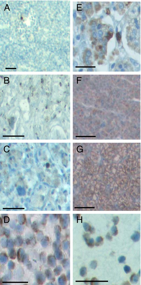
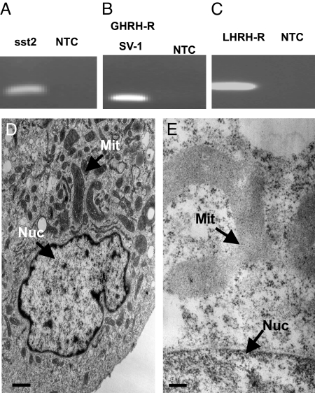
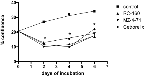
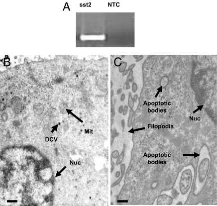
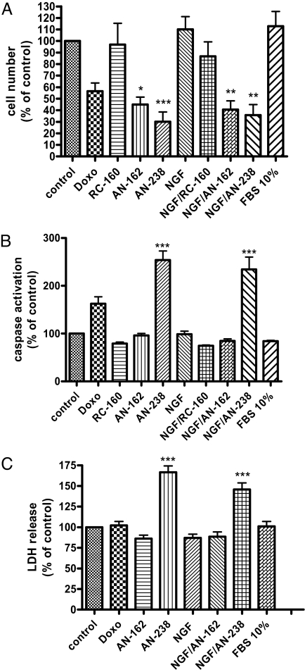
Peptide analogues targeting various neuropeptide receptors have been used effectively in cancer therapy. A hallmark of adrenocortical tumor formation is the aberrant expression of peptide receptors relating to uncontrolled cell proliferation and hormone overproduction. Our microarray results have also demonstrated a differential expression of neuropeptide hormone receptors in tumor subtypes of human pheochromocytoma. In light of these findings, we performed a comprehensive analysis of relevant receptors in both human adrenomedullary and adrenocortical tumors and tested the antiproliferative effects of peptide analogues targeting these receptors. Specifically, we examined the receptor expression of somatostatin-type-2 receptor, growth hormone-releasing hormone (GHRH) receptor or GHRH receptor splice variant-1 (SV-1) and luteinizing hormone-releasing hormone (LHRH) receptor at the mRNA and protein levels in normal human adrenal tissues, adrenocortical and adrenomedullary tumors, and cell lines. Cytotoxic derivatives of somatostatin AN-238 and, to a lesser extent, AN-162, reduced cell numbers of uninduced and NGF-induced adrenomedullary pheochromocytoma cells and adrenocortical cancer cells. Both the splice variant of GHRH receptor SV-1 and the LHRH receptor were also expressed in adrenocortical cancer cell lines but not in the pheochromocytoma cell line. The GHRH receptor antagonist MZ-4-71 and LHRH antagonist Cetrorelix both significantly reduced cell growth in the adrenocortical cancer cell line. In conclusion, the expression of receptors for somatostatin, GHRH, and LHRH in the normal human adrenal and in adrenal tumors, combined with the growth-inhibitory effects of the antitumor peptide analogues, may make possible improved treatment approaches to adrenal tumors.
Conflict of interest statement
The authors declare no conflict of interest.
Figures
Similar articles
-
Anti-tumor effects of peptide analogs targeting neuropeptide hormone receptors on mouse pheochromocytoma cells.Mol Cell Endocrinol. 2013 May 22;371(1-2):189-94. doi: 10.1016/j.mce.2012.12.011. Epub 2012 Dec 23. Mol Cell Endocrinol. 2013. PMID: 23267837 Free PMC article.
-
Therapy of experimental hepatic cancers with cytotoxic peptide analogs targeted to receptors for luteinizing hormone-releasing hormone, somatostatin or bombesin.Anticancer Drugs. 2008 Apr;19(4):349-58. doi: 10.1097/CAD.0b013e3282f9adce. Anticancer Drugs. 2008. PMID: 18454045
-
Receptors for luteinizing hormone releasing hormone expressed on human renal cell carcinomas can be used for targeted chemotherapy with cytotoxic luteinizing hormone releasing hormone analogues.Clin Cancer Res. 2005 Aug 1;11(15):5549-57. doi: 10.1158/1078-0432.CCR-04-2464. Clin Cancer Res. 2005. PMID: 16061872
-
New treatment approaches for prostate cancer based on peptide analogues.Eur Urol. 2008 May;53(5):890-900. doi: 10.1016/j.eururo.2007.12.021. Epub 2007 Dec 26. Eur Urol. 2008. PMID: 18201818 Review.
-
Targeting cytotoxic conjugates of somatostatin, luteinizing hormone-releasing hormone and bombesin to cancers expressing their receptors: a "smarter" chemotherapy.Curr Pharm Des. 2005;11(9):1167-80. doi: 10.2174/1381612053507594. Curr Pharm Des. 2005. PMID: 15853664 Review.
Cited by
-
Metastatic Pheochromocytoma and Paraganglioma: Somatostatin Receptor 2 Expression, Genetics, and Therapeutic Responses.J Clin Endocrinol Metab. 2023 Sep 18;108(10):2676-2685. doi: 10.1210/clinem/dgad166. J Clin Endocrinol Metab. 2023. PMID: 36946182 Free PMC article.
-
Epigenetic drugs in somatostatin type 2 receptor radionuclide theranostics and radiation transcriptomics in mouse pheochromocytoma models.Theranostics. 2023 Jan 1;13(1):278-294. doi: 10.7150/thno.77918. eCollection 2023. Theranostics. 2023. PMID: 36593963 Free PMC article.
-
THE ROLE OF TUMOR-SEEKING RADIOPHARMACEUTICALS IN THE DIAGNOSIS AND MANAGEMENT OF ADRENAL TUMORS.Acta Endocrinol (Buchar). 2020 Jul-Sep;16(3):316-323. doi: 10.4183/aeb.2020.316. Acta Endocrinol (Buchar). 2020. PMID: 33363653 Free PMC article.
-
Growth hormone-releasing hormone and its analogues in health and disease.Nat Rev Endocrinol. 2025 Mar;21(3):180-195. doi: 10.1038/s41574-024-01052-1. Epub 2024 Nov 13. Nat Rev Endocrinol. 2025. PMID: 39537825 Review.
-
Agonist of growth hormone-releasing hormone as a potential effector for survival and proliferation of pancreatic islets.Proc Natl Acad Sci U S A. 2010 Jul 13;107(28):12623-8. doi: 10.1073/pnas.1005098107. Epub 2010 Jun 28. Proc Natl Acad Sci U S A. 2010. PMID: 20616039 Free PMC article.
References
-
- Lacroix A, Ndiaye N, Tremblay J, Hamet P. Ectopic and abnormal hormone receptors in adrenal Cushing's syndrome. Endocr Rev. 2001;22:75–110. - PubMed
-
- Mazzuco TL, Chabre O, Feige JJ, Thomas M. Aberrant GPCR expression is a sufficient genetic event to trigger adrenocortical tumorigenesis. Mol Cell Endocrinol. 2007;265–266:23–28. - PubMed
-
- Willenberg HS, et al. Corticotropin-releasing hormone receptor expression on normal and tumorous human adrenocortical cells. Neuroendocrinology. 2005;82:274–281. - PubMed
-
- Willenberg HS, et al. Aberrant interleukin-1 receptors in a cortisol-secreting adrenal adenoma causing Cushing's syndrome. N Engl J Med. 1998;339:27–31. - PubMed
-
- Lampron A, et al. Regulation of aldosterone secretion by several aberrant receptors including for glucose-dependent insulinotropic peptide in a patient with an aldosteronoma. J Clin Endocrinol Metab. 2009;94:750–756. - PubMed
Publication types
MeSH terms
Substances
LinkOut - more resources
Full Text Sources
Other Literature Sources
Medical
