. 2009 Sep 15;106(37):15950-5.
doi: 10.1073/pnas.0900903106. Epub 2009 Aug 26.

Selective activation of the M1 muscarinic acetylcholine receptor achieved by allosteric potentiation

Affiliations

Selective activation of the M1 muscarinic acetylcholine receptor achieved by allosteric potentiation

Lei Ma et al. Proc Natl Acad Sci U S A. .

Erratum in

  • Proc Natl Acad Sci U S A. 2009 Oct 20;106(42):18040. Seager, Matthew [corrected to Seager, Matthew A]

Abstract

The forebrain cholinergic system promotes higher brain function in part by signaling through the M(1) muscarinic acetylcholine receptor (mAChR). During Alzheimer's disease (AD), these cholinergic neurons degenerate, therefore selectively activating M(1) receptors could improve cognitive function in these patients while avoiding unwanted peripheral responses associated with non-selective muscarinic agonists. We describe here benzyl quinolone carboxylic acid (BQCA), a highly selective allosteric potentiator of the M(1) mAChR. BQCA reduces the concentration of ACh required to activate M(1) up to 129-fold with an inflection point value of 845 nM. No potentiation, agonism, or antagonism activity on other mAChRs is observed up to 100 microM. Furthermore studies in M(1)(-/-) mice demonstrates that BQCA requires M(1) to promote inositol phosphate turnover in primary neurons and to increase c-fos and arc RNA expression and ERK phosphorylation in the brain. Radioligand-binding assays, molecular modeling, and site-directed mutagenesis experiments indicate that BQCA acts at an allosteric site involving residues Y179 and W400. BQCA reverses scopolamine-induced memory deficits in contextual fear conditioning, increases blood flow to the cerebral cortex, and increases wakefulness while reducing delta sleep. In contrast to M(1) allosteric agonists, which do not improve memory in scopolamine-challenged mice in contextual fear conditioning, BQCA induces beta-arrestin recruitment to M(1), suggesting a role for this signal transduction mechanism in the cholinergic modulation of memory. In summary, BQCA exploits an allosteric potentiation mechanism to provide selectivity for the M(1) receptor and represents a promising therapeutic strategy for cognitive disorders.


Conflict of interest statement

Conflict of interest statement: All authors were employed by Merck and Company, Inc. at the time of this study

Figures
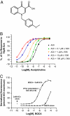
Fig. 1.
Fig. 1.
Potentiation Activity of BQCA. (A) Structure of BQCA. (B) Calcium mobilization measured by FLIPR in hM1-CHO cells induced by ACh (black line), or in the presence of BQCA at the indicated concentrations (colored lines). BQCA does not change the maximal response but causes a leftward shift in the ACh dose-response curve. Mean values from four replicate wells are plotted; data are representative of 12 independent experiments. (C) BQCA effects on calcium mobilization in hM1-CHO cells as measured by FLIPR when added at the indicated concentrations alone (bottom line) or in the presence of 3 nM ACh (top line). Effect of 3 nM ACh alone is plotted as single point, lower left corner. Each point is the mean of eight replicate determinations ± SEM and is representative of 225 independent experiments.
Fig. 2.
Fig. 2.
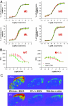
Selectivity of BQCA. (A) BQCA effects on calcium mobilization measured by FLIPR on M2-M5 mAChRs in CHO cells; legend as in Fig. 1B. Mean values from four replicate wells are plotted. To measure calcium mobilization by M2 and M4, which are Gi coupled and do not normally elicit a calcium response, a permissive recombinant G protein, Gqi5, was expressed in those cell lines. BQCA had no effect on the other human mAChRs at the highest concentrations tested (100 μM). Data shown are representative of >3 independent measurements. (B) IP1 measurements by HTRF in wild-type or M1−/− mouse primary neurons shows that ACh causes a dose-dependent decrease in counts, indicating increased IP1 levels and thus inositol phosphate metabolism, in wild-type and M1−/− neurons. Error bars, SEM. (n = four replicate wells) and the data are representative of four independent experiments. (C) In situ hybridization for c-Fos (Top) and arc (Bottom) RNAs in sagital sections of wild-type or M1−/− mice brains taken 1.5 h after oral dosing with 15 mg/kg BQCA or vehicle (saline).
Fig. 3.
Fig. 3.
BQCA is an allosteric potentiator. (A) Radioligand displacement assay using hM1-CHO membranes with unlabeled NMS (circles) and BQCA (triangles). Error bars, SEM (n = 4 replicate wells) and the data are representative of three experiments. (B) Radioligand binding assay measuring the displacement of NMS from hM1-CHO membranes by ACh ± BQCA. (C) [35]S-GTPγS bound to hM1-CHO membranes in the presence of ACh (squares) or increasing concentrations of BQCA. Error bars, SEM (n = four replicate wells); data are representative of three experiments. (D) Molecular modeling of the M1 extracellular region proximal to the ACh (red space-filling diagram in the center) binding site. A potential BQCA binding site was identified near amino acids Y179 and W400. (E) FLIPR data from Y179A and W400A mutations for ACh (black line) plus BQCA (colored lines). Legend and data format is as in Fig. 1B. (F) Amino acids flanking Y179 and W400 in human mAChRs.
Fig. 4.
Fig. 4.
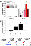
Physiological effects of BQCA. (A) Contextual fear conditioning. On day one animals received 0.3 mg/kg scopolamine ± BQCA administered IP at the indicated doses before being introduced to a novel environment and receiving two foot shocks. Twenty-four hours later animals (n = 12–16/group) were reintroduced to the environment and freezing measured by automated detection equipment. Shown is mean percent of time freezing (± SEM.). Data are representative of four experiments. *, Different from vehicle; #, different from scopolamine + vehicle (P < 0.05, Dunnet test). (B) Increased cerebral blood flow in anesthetized rats in response to BQCA. Data were averaged over 1 min at the indicated time points (A–C) and is expressed as mean percent change from baseline ± SEM. (n = 4 animals). *, Significantly different from baseline, P < 0.01, repeated measures ANOVA, t test.
Fig. 5.
Fig. 5.
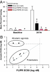
Allosteric agonists do not reverse scopolamine deficits in contextual fear conditioning or recruit β-arrestin. (A) Effects of TBPB at 10 and 30 mg/kg (mpk) in contextual fear conditioning. Shown is mean (n = 16/group) percent of time exhibiting freezing behavior + SEM *, Different from vehicle alone (P < 0.05, Dunnet test). Experiment was repeated twice with similar results. (B) TBPB and AC-42 (allosteric agonists, top circle) do not significantly recruit β-arrestin to hM1 compared to BQCA and 8 related allosteric potentiators (allosteric potentiators, bottom circle). Potencies (IP) for these compounds in calcium mobilization as measured by FLIPR (x axis) were compared to β-arrestin recruitment as measured by enzyme complementation (y axis). In both experiments, CHO cells stably expressing hM1 were used, for β-arrestin hM1 is fused to a portion of β-galactosidase while a complementary β-galactosidase fragment is fused to β-arrestin. Thus β-galactosidase activity is an indirect measure of β-arrestin recruitment. Values along the dashed lines indicate that no inflection point in the slope was observed at the highest concentrations tested (representative dose–response curves are in Fig. S8C). Note that the allosteric potentiators show a correlation between calcium mobilization and β-arrestin recruitment whereas for the allosteric agonists IP values were only measurable in calcium mobilization.

References

    1. Geula C. Abnormalities of neural circuitry in Alzheimer's disease: Hippocampus and cortical cholinergic innervation. Neurology. 1998;51:S18–29. - PubMed
    1. Hansen R, et al. Efficacy and safety of donepezil, galantamine, and rivastigmine for the treatment of Alzheimer's disease: A systematic review and meta-analysis. Clin Interv Aging. 2008;3:211–225. - PMC - PubMed
    1. Levey A. Muscarinic acetylcholine receptor expression in memory circuits: Implications for treatment of Alzheimer disease. Proc Natl Acad Sci USA. 1996;93:13541–13546. - PMC - PubMed
    1. Lanzafame A, Christopoulos A, Mitchelson F. Cellular signaling mechanisms for muscarinic acetylcholine receptors. Receptors Channels. 2003;9:241–260. - PubMed
    1. Felder C. Muscarinic acetylcholine receptors: Signal transduction through multiple effectors. FASEB J. 1995;9:619–625. - PubMed

MeSH terms