. 2009 Sep 15;106(37):15696-701.
doi: 10.1073/pnas.0811261106. Epub 2009 Aug 26.

The regulatory role of cell mechanics for migration of differentiating myeloid cells

Affiliations

The regulatory role of cell mechanics for migration of differentiating myeloid cells

Franziska Lautenschläger et al. Proc Natl Acad Sci U S A. .

Abstract

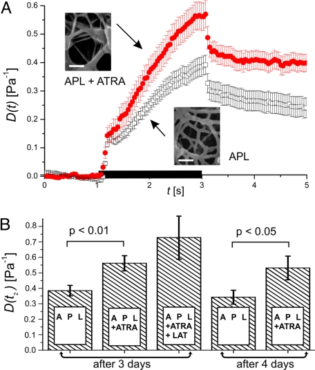
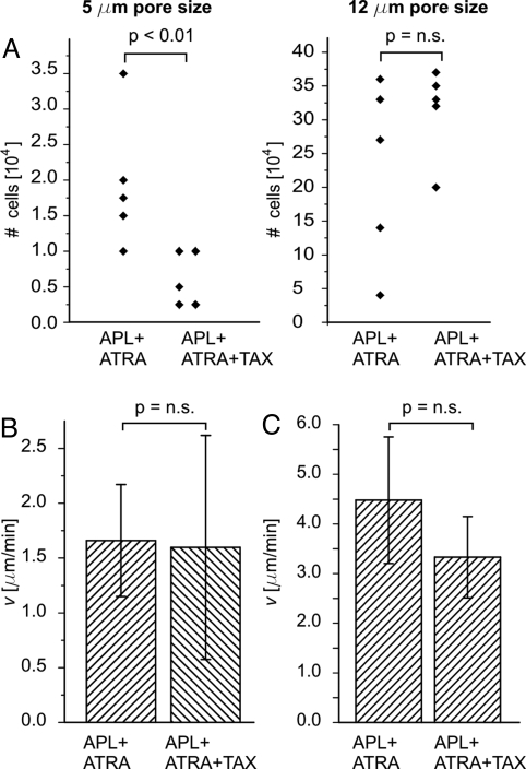
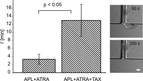
Migration of cells is important for tissue maintenance, immune response, and often altered in disease. While biochemical aspects, including cell adhesion, have been studied in detail, much less is known about the role of the mechanical properties of cells. Previous measurement methods rely on contact with artificial surfaces, which can convolute the results. Here, we used a non-contact, microfluidic optical stretcher to study cell mechanics, isolated from other parameters, in the context of tissue infiltration by acute promyelocytic leukemia (APL) cells, which occurs during differentiation therapy with retinoic acid. Compliance measurements of APL cells reveal a significant softening during differentiation, with the mechanical properties of differentiated cells resembling those of normal neutrophils. To interfere with the migratory ability acquired with the softening, differentiated APL cells were exposed to paclitaxel, which stabilizes microtubules. This treatment does not alter compliance but reduces cell relaxation after cessation of mechanical stress six-fold, congruent with a significant reduction of motility. Our observations imply that the dynamical remodeling of cell shape required for tissue infiltration can be frustrated by stiffening the microtubular system. This link between the cytoskeleton, cell mechanics, and motility suggests treatment options for pathologies relying on migration of cells, notably cancer metastasis.


Conflict of interest statement

Conflict of interest statement: J.G. holds a patent on the optical stretcher technique and consults on its potential applications.

Figures

Fig. 1.
Fig. 1.
Principle and setup of a microfluidic optical stretcher (μOS). (A) Two-counter propagating NIR laser beams (λ = 1,064 nm) emanating from the cores of single-mode optical fibers are used to trap (P = 0.1 W each beam) and deform (P = 1 W each beam) single cells. Cells are deformed by the forces arising from the momentum transfer of light to the surface of the cell due to the change in refractive index. (B) Experimental setup. The flow of the cell suspension between two reservoirs (Res 1 and Res 2) is controlled by a hydrostatic pressure differential. The phase contrast image shows the microfluidic flowchamber with a cell trapped inside the glass microcapillary. (C) Phase-contrast images of APL cell being optically trapped (left) and stretched (middle) in the μOS. Optically induced surface stress causes a deformation of the cell along the optical axis. (Right) overlay of the two other images and definition of the strain, γ(t) = Δr(t)/r0. (Scale bar, 5 μm.)
Fig. 2.
Fig. 2.
Change of APL cell compliance in response to ATRA. (A) ATRA differentiated APL cells (filled circles; n = 44) are significantly more compliant than controls (open squares; n = 42), mean ± SEM. The black bar on the time axis indicates the application of stress. Insets depict electron microscopy images of the subcortical actin filament network. (Scale bars, 100 nm.) (B) Deformability at the end of stress application, mean ± SEM. There was no significant difference between APL cells exposed to ATRA for 3 and 4 days. A higher deformability could be measured after depolymerizing F-actin with latrunculin A (LAT).
Fig. 3.
Fig. 3.
Relaxation behavior of ATRA differentiated APL cells before and after exposure to paclitaxel. The relaxation of differentiated APL cells after cessation of stress is decreased when treated with paclitaxel (TAX). (filled circles) APL cells treated with ATRA (n = 44), (open squares) APL cells treated with ATRA and paclitaxel (n = 56). The duration of stress application is indicated by the black bar on the time axis. Data show mean ± SEM. Insets show fluorescence images of the microtubule organization in both groups of cells.
Fig. 4.
Fig. 4.
Migration of differentiated APL cells. (A) Chemotaxis through small pores. Paclitaxel significantly impedes the migration within 3 h of differentiated APL cells through 5-μm large pores (left). There is no statistically significant difference for 12-μm pores (right). Both experiments were performed five times. No change in chemotactic speed of differentiated APL cells could be observed after treatment with paclitaxel on 2D surfaces (B) or in 3D microchannels of 5–7-μm width (C). Data show mean ± SEM.
Fig. 5.
Fig. 5.
Pore entry as rate-limiting step. Paclitaxel significantly increases the entry time of differentiated APL cells into 3D channels (ntotal = 11). Data are mean ± SEM. Insets show a differentiated APL cell before and after entering the microfluidic channel (7-μm width). (Scale bar, 10 μm.)

References

    1. Dormann D, Weijer CJ. Imaging of cell migration. EMBO J. 2006;25:3480–3493. - PMC - PubMed
    1. Duffy MJ, McGowan PM, Gallagher WM. Cancer invasion and metastasis: Changing views. J Pathol. 2008;214:283–293. - PubMed
    1. Even-Ram S, Yamada KM. Cell migration in 3D matrix. Curr Opin Cell Biol. 2005;17:524–532. - PubMed
    1. Wolf K, et al. Compensation mechanism in tumor cell migration: mesenchymal-amoeboid transition after blocking of pericellular proteolysis. J Cell Bio. 2003;160:267–277. - PMC - PubMed
    1. Friedl P, Weigelin B. Interstitial leukocyte migration and immune function. Nature Immunol. 2008;9:960–969. - PubMed

Publication types

MeSH terms