

A role for galanin in human and experimental inflammatory demyelination
- PMID: 19717462
- PMCID: PMC2732706
- DOI: 10.1073/pnas.0903360106
A role for galanin in human and experimental inflammatory demyelination
Abstract
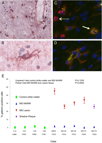
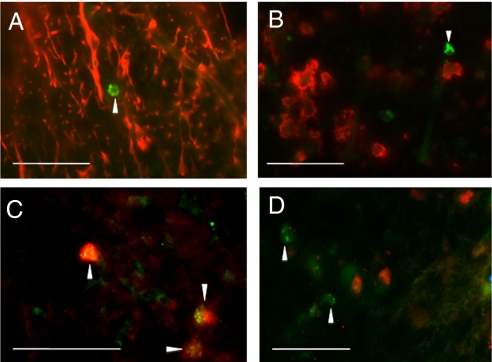
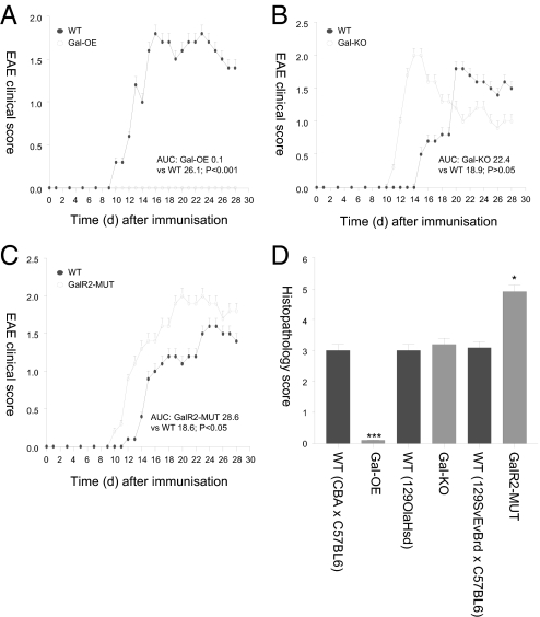
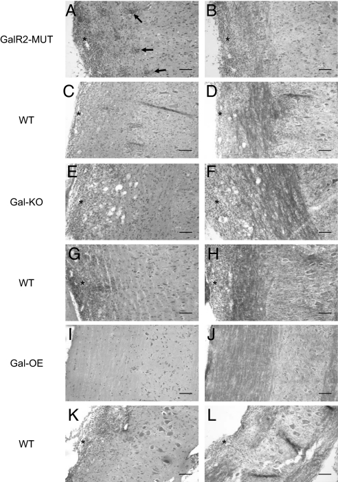
The neuropeptide galanin is widely expressed by many differing subsets of neurons in the nervous system. There is a marked upregulation in the levels of the peptide in a variety of nerve injury models and in the basal forebrain of humans with Alzheimer's disease. Here we demonstrate that galanin expression is specifically and markedly upregulated in microglia both in multiple sclerosis (MS) lesions and shadow plaques. Galanin expression is also upregulated in the experimental autoimmune encephalomyelitis (EAE) model of MS, although solely in oligodendrocytes. To study whether the observed increase in expression of galanin in inflammatory demyelination might modulate disease activity, we applied the EAE model to a panel of galanin transgenic lines. Over-expression of galanin in transgenic mice (Gal-OE) abolishes disease in the EAE model, whilst loss-of-function mutations in galanin or galanin receptor-2 (GalR2) increase disease severity. The pronounced effects of altered endogenous galanin or GalR2 expression on EAE disease activity may reflect a direct neuroprotective effect of the neuropeptide via activation of GalR2, similar to that previously described in a number of neuronal injury paradigms. Irrespective of the mechanism(s) by which galanin alters EAE disease activity, our findings imply that galanin/GalR2 agonists may have future therapeutic implications for MS.
Conflict of interest statement
The authors declare no conflict of interest.
Figures
Similar articles
-
Detection of galanin receptors in the spinal cord in experimental autoimmune encephalomyelitis.Biomed Pap Med Fac Univ Palacky Olomouc Czech Repub. 2023 Mar;167(1):36-42. doi: 10.5507/bp.2022.005. Epub 2022 Feb 11. Biomed Pap Med Fac Univ Palacky Olomouc Czech Repub. 2023. PMID: 35147137
-
Ectopic galanin expression and normal galanin receptor 2 and galanin receptor 3 mRNA levels in the forebrain of galanin transgenic mice.Neuroscience. 2005;133(2):371-80. doi: 10.1016/j.neuroscience.2005.01.068. Neuroscience. 2005. PMID: 15885921
-
Galanin acts as a trophic factor to the central and peripheral nervous systems.Exp Suppl. 2010;102:25-38. doi: 10.1007/978-3-0346-0228-0_3. Exp Suppl. 2010. PMID: 21299059 Review.
-
Activation of the galanin receptor 2 (GalR2) protects the hippocampus from neuronal damage.J Neurochem. 2007 Feb;100(3):780-9. doi: 10.1111/j.1471-4159.2006.04239.x. J Neurochem. 2007. PMID: 17263796 Free PMC article.
-
Galanin acts as a trophic factor to the central and peripheral nervous systems.Cell Mol Life Sci. 2008 Jun;65(12):1806-12. doi: 10.1007/s00018-008-8154-7. Cell Mol Life Sci. 2008. PMID: 18500646 Free PMC article. Review.
Cited by
-
Generating orally active galanin analogues with analgesic activities.ChemMedChem. 2012 May;7(5):903-9. doi: 10.1002/cmdc.201100574. Epub 2012 Feb 28. ChemMedChem. 2012. PMID: 22374865 Free PMC article.
-
Intranasal Delivery of a Methyllanthionine-Stabilized Galanin Receptor-2-Selective Agonist Reduces Acute Food Intake.Neurotherapeutics. 2021 Oct;18(4):2737-2752. doi: 10.1007/s13311-021-01155-x. Epub 2021 Dec 2. Neurotherapeutics. 2021. PMID: 34859381 Free PMC article.
-
Prolonged stimulation of a brainstem raphe region attenuates experimental autoimmune encephalomyelitis.Neuroscience. 2017 Mar 27;346:395-402. doi: 10.1016/j.neuroscience.2017.01.037. Epub 2017 Jan 29. Neuroscience. 2017. PMID: 28147248 Free PMC article.
-
Neuroimmunomodulation by gut bacteria: Focus on inflammatory bowel diseases.World J Gastrointest Pathophysiol. 2021 May 22;12(3):25-39. doi: 10.4291/wjgp.v12.i3.25. World J Gastrointest Pathophysiol. 2021. PMID: 34084590 Free PMC article. Review.
-
Detection of galanin receptors in the spinal cord in experimental autoimmune encephalomyelitis.Biomed Pap Med Fac Univ Palacky Olomouc Czech Repub. 2023 Mar;167(1):36-42. doi: 10.5507/bp.2022.005. Epub 2022 Feb 11. Biomed Pap Med Fac Univ Palacky Olomouc Czech Repub. 2023. PMID: 35147137
References
-
- Tatemoto K, Rokaeus A, Jornvall H, McDonald TJ, Mutt V. Galanin - A novel biologically active peptide from porcine intestine. FEBS Lett. 1983;164:124–128. - PubMed
-
- Ch'ng JL, et al. Distribution of galanin immunoreactivity in the central nervous system and the responses of galanin-containing neuronal pathways to injury. Neuroscience. 1985;16:343–354. - PubMed
-
- Melander T, Hokfelt T, Rokaeus A. Distribution of galaninlike immunoreactivity in the rat central nervous system. J Comp Neurol. 1986;248:475–517. - PubMed
-
- Rokaeus A, et al. A galanin-like peptide in the central nervous system and intestine of the rat. Neurosci Lett. 1984;47:161–166. - PubMed
-
- Skofitsch G, Jacobowitz DM. Galanin-like immunoreactivity in capsaicin sensitive sensory neurons and ganglia. Brain Res Bull. 1985;15:191–195. - PubMed
Publication types
MeSH terms
Substances
Grants and funding
LinkOut - more resources
Full Text Sources
Other Literature Sources
Medical
Molecular Biology Databases
