. 2009 Sep 15;106(37):15750-5.
doi: 10.1073/pnas.0908332106. Epub 2009 Aug 26.

Mammalian MagT1 and TUSC3 are required for cellular magnesium uptake and vertebrate embryonic development

Affiliations

Mammalian MagT1 and TUSC3 are required for cellular magnesium uptake and vertebrate embryonic development

Hao Zhou et al. Proc Natl Acad Sci U S A. .

Abstract

Magnesium (Mg(2+)) is the second most abundant cation in cells, yet relatively few mechanisms have been identified that regulate cellular levels of this ion. The most clearly identified Mg(2+) transporters are in bacteria and yeast. Here, we use a yeast complementary screen to identify two mammalian genes, MagT1 and TUSC3, as major mechanisms of Mg(2+) influx. MagT1 is universally expressed in all human tissues and its expression level is up-regulated in low extracellular Mg(2+). Knockdown of either MagT1 or TUSC3 protein significantly lowers the total and free intracellular Mg(2+) concentrations in mammalian cell lines. Morpholino knockdown of MagT1 and TUSC3 protein expression in zebrafish embryos results in early developmental arrest; excess Mg(2+) or supplementation with mammalian mRNAs can rescue the effects. We conclude that MagT1 and TUSC3 are indispensable members of the vertebrate plasma membrane Mg(2+) transport system.


Conflict of interest statement

The authors declare no conflict of interest.

Figures
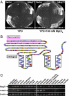
Fig. 1.
Fig. 1.
MagT1 and TUSC3 complement the yeast ALR1 Mg2+ transporter-deficient strain. (A) Yeast complementation assay. MagT1 and two splicing isoforms of TUSC3 were subcloned into the yeast p413GPD expression vector and the alr1Δ strain transformed. Yeasts were streaked on either YPD or YPD plates supplemented with 100 mM MgCl2, and grown for 2 days at 30 °C. (B) Predicted MagT1 secondary structure with the positions of transmembrane (TM) helices and signal peptide indicated. (C) RT-PCR of mRNA from human tissues as indicated.
Fig. 2.
Fig. 2.
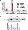
MagT1 is up-regulated in low Mg2+. (A) Quantitative RT-PCR of total RNA from HEK 293T cells grown in either standard DMEM, or media with the indicated [Mg2+] for 1 or 2 days. Samples were measured in triplicate, and β-actin levels were used as internal controls. Error bars indicate SEM, and asterisks indicate P < 0.01. (B) Western blots on cells grown in the media indicated in (A) using polyclonal antibody against human MagT1. The relative intensity of the Western signal is shown (Bottom).
Fig. 3.
Fig. 3.
MagT1 is located on the cell surface. (A) Biotinylation of overexpressed MagT1 protein. HEK 293T cells were transfected with HA-tagged MagT1, grown for 24 h, and surface proteins labeled by biotinylation. Total cell lysate and streptavidin-purified portions were loaded onto SDS/PAGE gels, and Western blots were performed with α-HA, α-tubulin, or α-Na, K-ATPase antibodies. (B) Biotinylation of native MagT1 protein in untransfected HEK 293T cells; native MagT1 protein was detected by α-MagT1 polyclonal antibody. (C) Immunostaining of transfected MagT1-HA protein in HEK 293T cells. Permeabilized (0.2% Triton X-100), or nonpermeabilized cell immunostaining with α-HA antibody are compared. (Scale bar, 10 μm.)
Fig. 4.
Fig. 4.
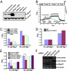
MagT1 and TUSC3 are required for cellular Mg2+ uptake. (A) RNA interference and overexpression of MagT1. HEK 293T cells were transfected with three different siRNAs or MagT1 cDNA and grown for 2 days before Western blotting total cell lysates using polyclonal α-hMagT1 and α-tubulin antibodies. (B) Representative traces of the Mg2+ assay in MagT1 knockdown cells. HEK 293T cells were loaded with 20 μM KMG104-AM (45 min, 37 °C), and fluorescence changes monitored in differing [Mg2+]. One molar Mg2+ was added at the end of the experiment. (C) Quantification of traces in (B), with peak fluorescence levels shown for each [Mg2+] treatment; error bars indicate SEM; asterisks indicate P < 0.01. (D) Mg2+ uptake assay on MagT1 and TUSC3 overexpressing cells. HEK 293T cells were cotransfected with MagT1 and TUSC3 and incubated for 24 h before the assay. (E) Mg2+ uptake assay in Jurkat cells with potential Mg2+ transporters (MagT1, SLC41A1, SLC41A2) knocked down. Each of the indicated siRNAs was transfected into Jurkat cells via electroporation and cells were incubated for 2 days before the assay. (F) RT-PCR on total RNA from the Jurkat cells in (E).
Fig. 5.
Fig. 5.
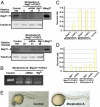
MagT1 and TUSC3 are required for zebrafish embryonic development. (A) Western blots of zebrafish embryo total lysates. Fertilized eggs were injected by one of two combinations of morpholinos against both MagT1 and TUSC3 (Morpholino A, Morpholino B; 100 μM each in final concentration) or buffer alone (Control). Embryos were collected after 30 h, homogenized, lysed, and anti-MagT1 and -tubulin antibodies used to recognize zebrafish proteins. Lysate from HEK cells with stable MagT1 expression (hMagT1) served as control. Sets of the same eggs were also coinjected with either 100 ng/μL of human MagT1 and TUSC3 mRNA, or 10 mM MgCl2, as indicated. The ratio between the MagT1 and tubulin signals was calculated and normalized to the controls. (B) RT-PCR of total RNA from the embryos injected with Morpholino B, showing effects on splicing. Eggs were injected as in (A), with the Morpholino B set. (C) Embryo survival rates after single morpholino injection. 48 h after injections with 200 μM each of the morpholinos indicated, survival rates were calculated as the ratio between total hatched larvae and total eggs injected. (D) Double morpholino injections and rescue by coinjection with Mg2+ and human MagT1 and TUSC3 mRNA. Eggs were injected with a combination of morpholinos against MagT1 and TUSC3 as in (A and B), and survival rates were calculated as in C. For C and D, 40–70 embryos were counted for each injection. Control, mock injection with buffer. (E) Zebrafish development is arrested after morpholino treatment. Fertilized eggs were injected with a combination of morpholino A against MagT1 and TUSC3 (100 μM each in final concentration) or buffer alone. Images: 30 h stage.

Similar articles

Cited by

References

    1. Romani A. Regulation of magnesium homeostasis and transport in mammalian cells. Arch Biochem Biophys. 2007;458:90–102. - PubMed
    1. Fatholahi M, LaNoue K, Romani A, Scarpa A. Relationship between total and free cellular Mg(2+) during metabolic stimulation of rat cardiac myocytes and perfused hearts. Arch Biochem Biophys. 2000;374:395–401. - PubMed
    1. Harman AW, Nieminen AL, Lemasters JJ, Herman B. Cytosolic free magnesium, ATP and blebbing during chemical hypoxia in cultured rat hepatocytes. Biochem Biophys Res Commun. 1990;170:477–483. - PubMed
    1. Raju B, Murphy E, Levy LA, Hall RD, London RE. A fluorescent indicator for measuring cytosolic free magnesium. Am J Physiol. 1989;256:C540–C548. - PubMed
    1. Tashiro M, Konishi M. Basal intracellular free Mg2+ concentration in smooth muscle cells of guinea pig tenia cecum: Intracellular calibration of the fluorescent indicator furaptra. Biophys J. 1997;73:3358–3370. - PMC - PubMed

Publication types

MeSH terms