. 2009 Nov;191(21):6482-8.
doi: 10.1128/JB.00965-09. Epub 2009 Aug 28.

Two essential arginine residues in the T components of energy-coupling factor transporters

Affiliations

Two essential arginine residues in the T components of energy-coupling factor transporters

Olivia Neubauer et al. J Bacteriol. 2009 Nov.

Abstract

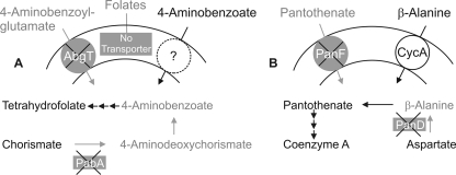
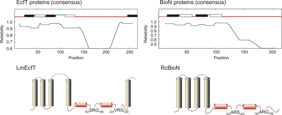
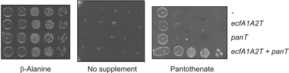
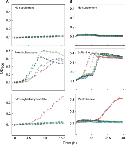
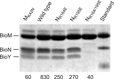
Energy-coupling factor (ECF) transporters, a recently discovered class of importers of micronutrients, are composed of a substrate-specific transmembrane component (S component) and a conserved energy-coupling module consisting of a transmembrane protein (T component) and pairs of ABC ATPases (A proteins). Based on utilization of a dedicated (subclass I) or shared (subclass II) energy-coupling module, ECF systems fall into two subclasses. The T components are the least-characterized proteins of ECF importers, and their function is essentially unknown. Using RcBioN and LmEcfT, the T units of the subclass I biotin transporter (RcBioMNY) of a gram-negative bacterium and of the subclass II folate, pantothenate, and riboflavin transporters of a lactic acid bacterium, respectively, we analyzed the role of two strongly conserved short motifs, each containing an arginine residue. Individual replacement of the two Arg residues in RcBioN reduced ATPase activity, an indicator of the transporter function, by two-thirds without affecting the modular assembly of the RcBioMNY complex. A double Arg-to-Glu replacement destroyed the complex and abolished ATPase activity. The corresponding single mutation in motif II of LmEcfT, as well as a double mutation, led to loss of the T unit from the subclass II ECF transporters and inactivated these systems. A single Arg-to-Glu replacement in motif I, however, abolished vitamin uptake activity without affecting assembly of the modules. Our results indicate that the conserved motif I in T components is essential for intramolecular signaling and, in cooperation with motif II, for subunit assembly of modular ECF transporters.


Figures

FIG. 1.
FIG. 1.
Phenotypes of vitamin synthesis- and vitamin transport-deficient E. coli strains. (A) The pabA abgT strain (kindly provided by A. Eudes and A. D. Hanson, Gainesville, FL) lacks the transporter for the folate catabolite 4-aminobenzoyl-glutamate and cannot produce the folate precursors 4-aminodeoxychorismate and 4-aminobenzoate. It can take up exogenous 4-aminobenzoate by an unknown transporter. E. coli K-12 naturally lacks transport systems for folates. (B) E. coli DV1 (panD panF) lacks the sodium/pantothenate symporter and is unable to produce the pantothenate precursor β-alanine. It can utilize exogenous β-alanine by means of the CycA amino acid transporter (15).
FIG. 2.
FIG. 2.
Topology of T components. (Upper panels) The consensus amino acid sequences of 23 EcfT proteins from lactobacteria (left panel) and 23 BioN proteins from gram-negative bacteria (right panel) were calculated with CLUSTALW. The consensus sequences were analyzed by TOPCONS based on the SCAMPI, PRO-TMHMM, PRODIV-TMHMM, and OCTOPUS algorithms. Predicted transmembrane helices are indicated by solid (in-to-out orientation) and open (out-to-in orientation) bars. Only the helices with a TOPCONS reliability score greater than 0.6 were considered. Red and blue indicate cytoplasmic and extracytoplasmic locations, respectively. (Lower panels) Topological models of the L. mesenteroides EcfT (LmEcfT) and R. capsulatus BioN (RcBioN) proteins based on the consensus profiles. The conserved signatures with “ARG” as the consensus sequence are located adjacent to helical regions with various hydrophobicities in individual T components. The colors indicate cytoplasmic and extracytoplasmic locations as described above.
FIG. 3.
FIG. 3.
Assay of pantothenate uptake activity. E. coli strain DV1 (see the text and Fig. 1B for the genotype and phenotype, respectively) containing an empty vector (−) or expressing the L. mesenteroides genes indicated on the right was pregrown in β-alanine-containing mineral salts medium. Cultures were harvested and washed in sterile buffer. Aliquots of 10-fold serial dilutions were spotted onto mineral salts agar plates containing 1 mM IPTG and either no additional supplement, 1 μM β-alanine, or 1 μM Ca2+-pantothenate.
FIG. 4.
FIG. 4.
Mutations in EcfT abolish folate and pantothenate uptake. (A) The E. coli pabA abgT strain containing pACYC-RIL, pLmFolT, and a variant of pLmEcfAAT with a wild-type (red circles), R184E (blue circles), R225E (black circles), or R184/225R (green circles) ecfT allele were grown in minimal medium containing no supplement, 10 μM 4-aminobenzoate, or 10 μM 5-formyl-tetrahydrofolate. (B) E. coli DV1 (panD panF) harboring pLacI-RARE2, pLmPanT, and variants of pLmEcfAAT as indicated above was grown in unsupplemented minimal medium or in medium containing 1 μM β-alanine or 1 μM Ca2+-pantothenate. All cultures contained 0.5 mM IPTG and were grown in duplicate in microtiter plates at 37°C. Growth was monitored by determining the optical density at 600 nm (OD600) and is expressed as the mean of duplicate results. See Fig. 1 for the strain phenotypes.
FIG. 5.
FIG. 5.
Complexes of EcfA1A2T with FolT, PanT, and RibU. E. coli BL21 containing pLacI-RARE2 was used as the host. The EcfA1A2T module with wild-type (WT) and mutant (R184E, R225E, R184/225E) ecfT genes was produced from a plasmid (resulting in deca-His-tagged EcfA1, untagged EcfA2, and FLAG-tagged EcfT) in the presence of another plasmid encoding FLAG-tagged FolT, PanT, or RibU. Membranes of the recombinants were isolated and solubilized with dodecyl-β-d-maltoside. Solubilized material was chromatographed on immobilized Ni-NTA. Enriched proteins were separated by SDS-PAGE and blotted onto nitrocellulose membranes. The membranes were probed with monoclonal anti-oligo-His or anti-FLAG antibody-alkaline phosphatase conjugates. The marker proteins (left lane in each panel) had molecular masses of (from top to bottom) 37, 25, and 20 kDa.
FIG. 6.
FIG. 6.
Consequences of replacements in BioN for complex formation and ATPase activity. BioMNY complexes were purified using the His tag on BioM. Two micrograms of protein was subjected to SDS-PAGE, and the gel was stained with Coomassie blue. The molecular masses of the standard proteins are 25, 20, and 15 kDa. The number below each lane represents the ATPase activity of the purified sample, expressed in nmol Pi min−1 (mg protein)−1. The BioMK42NNY complex (left lane) with inactivated ATPase domains served as a negative control.

References

    1. Baba, T., T. Ara, M. Hasegawa, Y. Takai, Y. Okumura, M. Baba, K. A. Datsenko, M. Tomita, B. L. Wanner, and H. Mori. 2006. Construction of Escherichia coli K-12 in-frame, single-gene knockout mutants: the Keio collection. Mol. Syst. Biol. 2:2006.0008. - PMC - PubMed
    1. Bernsel, A., H. Viklund, J. Falk, E. Lindahl, G. von Heijne, and A. Elofsson. 2008. Prediction of membrane-protein topology from first principles. Proc. Natl. Acad. Sci. USA 105:7177-7181. - PMC - PubMed
    1. Bernsel, A., H. Viklund, A. Hennerdal, and A. Elofsson. 2009. TOPCONS: consensus prediction of membrane protein topology. Nucleic Acids Res. 37:W465-W468. - PMC - PubMed
    1. Burgess, C. M., D. J. Slotboom, E. R. Geertsma, R. H. Duurkens, B. Poolman, and D. van Sinderen. 2006. The riboflavin transporter RibU in Lactococcus lactis: molecular characterization of gene expression and the transport mechanism. J. Bacteriol. 188:2752-2760. - PMC - PubMed
    1. Davidson, A. L., E. Dassa, C. Orelle, and J. Chen. 2008. Structure, function, and evolution of bacterial ATP-binding cassette systems. Microbiol. Mol. Biol. Rev. 72:317-364. - PMC - PubMed

Publication types

MeSH terms

LinkOut - more resources