. 2009 Aug;21(8):2220-36.
doi: 10.1105/tpc.109.065730. Epub 2009 Aug 28.

The Arabidopsis CORONATINE INSENSITIVE1 protein is a jasmonate receptor

Affiliations

The Arabidopsis CORONATINE INSENSITIVE1 protein is a jasmonate receptor

Jianbin Yan et al. Plant Cell. 2009 Aug.

Abstract

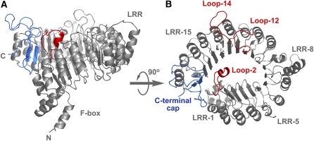
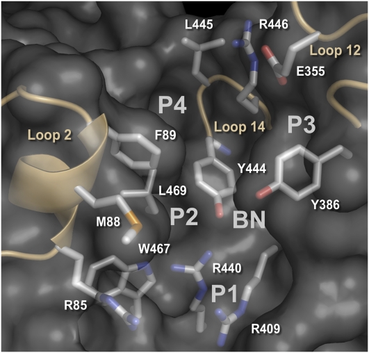
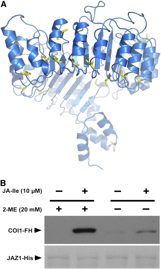
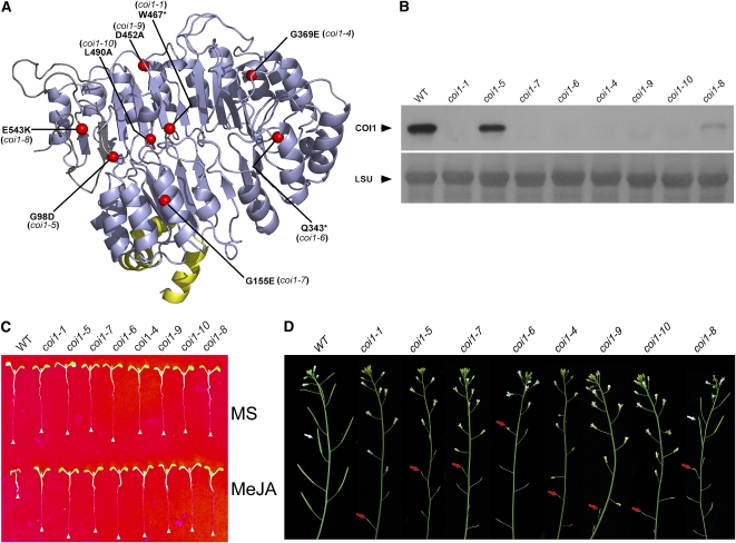
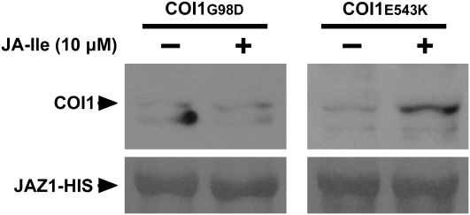
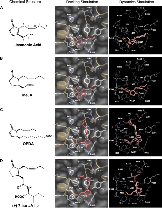
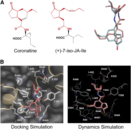
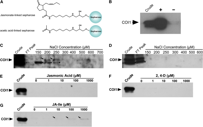
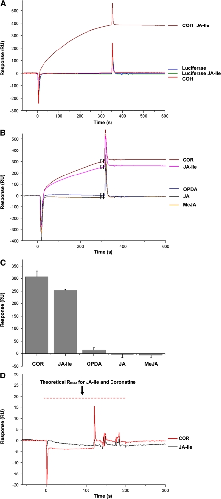
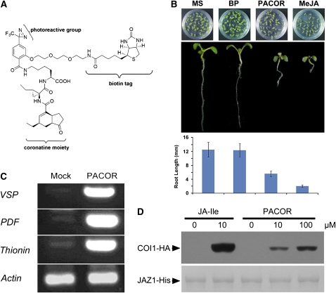
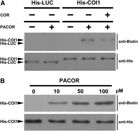
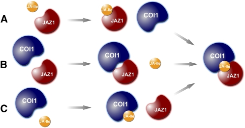
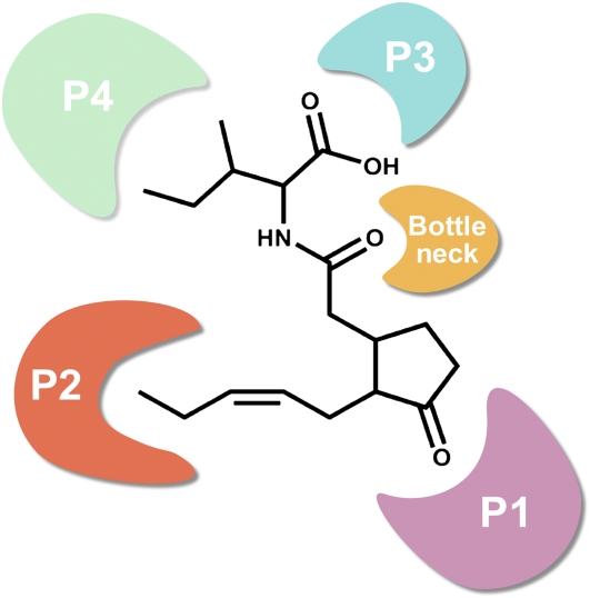
Jasmonates play a number of diverse roles in plant defense and development. CORONATINE INSENSITIVE1 (COI1), an F-box protein essential for all the jasmonate responses, interacts with multiple proteins to form the SCF(COI1) E3 ubiquitin ligase complex and recruits jasmonate ZIM-domain (JAZ) proteins for degradation by the 26S proteasome. To determine which protein directly binds to jasmonoyl-isoleucine (JA-Ile)/coronatine (COR) and serves as a receptor for jasmonate, we built a high-quality structural model of COI1 and performed molecular modeling of COI1-jasmonate interactions. Our results imply that COI1 has the structural traits for binding JA-Ile or COR. The direct binding of these molecules with COI1 was further examined using a combination of molecular and biochemical approaches. First, we used the immobilized jasmonate approach to show that the COI1 protein in crude leaf extracts can bind to the jasmonate moiety of JA-Ile. Second, we employed surface plasmon resonance technology with purified COI1 and JAZ1 protein to reveal the interaction among COI1, JA-Ile, and JAZ1. Finally, we used the photoaffinity labeling technology to show the direct binding of COR with purified insect-expressed COI1. Taken together, these results demonstrate that COI1 directly binds to JA-Ile and COR and serves as a receptor for jasmonate.


Figures

Figure 1.
Figure 1.
Structural Model of COI1. Two views of the structural model of COI1 are shown as ribbon diagrams. The F-box and LRR domains of COI1 are labeled and shown in gray. The C-terminal cap is shown in blue. The 18 LRR motifs are numbered. The three surface loops in LRR-2, LRR-12, and LRR-14 are shown in red and labeled loop-2, loop-12, and loop-14.
Figure 2.
Figure 2.
An Overall View of the Surface Pocket in COI1. Three long loops (loop 2, loop 12, and loop 14) and the solenoid inner surface of COI1 form a surface pocket that can be divided into four distinct pockets (P1, P2, P3, and P4) and one bottleneck (BN) region based on surface properties. The P1 pocket mainly includes basic residues (Arg-409, Arg-440, and Arg-85), and the P2 pocket is encircled by hydrophobic residues (Met-88, Phe-89, and Trp-467). The P3 pocket mainly contains hydrophilic residues (Glu-355 and Arg-446), while the P4 pocket mainly contains hydrophobic residues (Leu-445 and Leu-469). The channel connecting the four pockets is occupied by the bottleneck region, which contains Tyr-386 and Tyr-444. The residues in the surface pocket are shown as green stick models. Loop 2, loop 12, and loop 14 are shown in yellow.
Figure 3.
Figure 3.
The Reducing Environment Is Important for the JA-Ile–Induced Interaction between COI1 and JAZ1. (A) A view of the Cys residues in the COI1 structural model. The structural model of COI1 is shown as a ribbon diagram. The Cys residues of COI1 are shown as yellow stick models. All the sulfhydryl groups of Cys residues are in a reduced form. (B) Pull-down reactions were performed using recombinant JAZ1-His and total plant extracts prepared from coi1-1 seedlings transgenic for HA-tagged COI1 (COI1-FH). The reactions were performed at 4°C for 1 h with or without 20 mM 2-mercaptoethanol at the indicated JA-Ile concentrations. Protein bound to the Ni-NTA resin was washed, separated on SDS-PAGE, and immunoblotted with an anti-HA antibody (top panel). The polyvinylidene fluoride (PVDF) membrane was stained with Memstain (Applygen Technologies) to visualize the recovery of JAZ1 by the Ni-NTA affinity resin (bottom panel).
Figure 4.
Figure 4.
Mutations in the LRR Domain Disrupt COI1 Stability in Vivo. (A) A view of the mutant sites in the COI1. The structural model of COI1 is shown as a ribbon diagram. The F-box, LRR domain, and C-terminal domain of COI1 are colored yellow, light blue, and gray, respectively. The mutant sites are labeled. (B) Determination of COI1 protein levels in coi1 mutant plants. The proteins were extracted from leaves and subjected to immunoblot analyses using COI1 antiserum. The PVDF membrane was stained with Memstain to visualize equal loading. LSU, large subunit of ribulose-1, 5-bisphosphate carboxylase/oxygenase. (C) Phenotype of 10-d-old seedlings grown on Murashige and Skoog (MS) medium or MS with 25 μM MeJA. The white triangles indicate root tips. (D) The coi1-8 displays a partial fertility phenotype, while the other mutants, like coi1-1, are male sterile. The red arrows indicate sterile siliques, and the white arrows indicate fertile siliques.
Figure 5.
Figure 5.
The G98D Mutation in COI1 Disrupts the JA-Ile–Dependent Interaction between COI1 and JAZ1. Pull-down assays used recombinant JAZ1-His protein and extracts from coi1-5 and coi1-8 mutants. The reactions were performed at 4°C for 1 h in the presence or absence of 10 μM JA-Ile. The recovery of COI1 was detected with an anti-COI1 antiserum (top panel). The PVDF membrane was stained with Memstain to visualize the recovery of JAZ1 by the Ni-NTA affinity resin (bottom panel).
Figure 6.
Figure 6.
JA-Ile Could Fit within the Surface Pocket of COI1. Molecular modeling of the interaction between COI1 and JA (A), MeJA (B), OPDA (C), or (+)-7-iso-JA-Ile (D). Left panel: chemical structures of the JAs. Middle panel: the pose with the highest GoldScore fitness value in the molecular docking simulation. The JAs are shown as red sticks. The surface pocket of COI1 is shown in gray. Right panel: superposition of representative frames of the restricted molecular dynamics simulation. Frames at the early, intermediate, and late stages were extracted and superimposed. The JAs are shown as pink sticks, and their interacting residues are shown as white lines. Polar contacts are shown as yellow dotted lines.
Figure 7.
Figure 7.
The Proposed Mechanism of COR Binding to COI1. (A) The similarity between (+)-7-iso-JA-Ile and COR. Chemical structures of COR (left) and (+)-7-iso-JA-Ile (middle) are shown. The groups of (+)-7-iso-JA-Ile or COR that contribute to the binding with the surface pocket of COI1 are colored red. The optimized binding conformation of (+)-7-iso-JA-Ile and COR was extracted and superimposed to show the identity of the binding conformations between (+)-7-iso-JA-Ile and COR in the surface pocket of COI1 (right). Carbon atoms in (+)-7-iso-JA-Ile are represented in red, while those in COR are in cyan. (B) Molecular modeling of the interaction between COI1 and COR. Left panel: the pose with the highest GoldScore fitness value in the molecular docking simulation. COR is shown in red sticks. The surface pocket of COI1 is shown in gray. Right panel: superposition of representative frames of the restricted molecular dynamics simulation. Frames at the early, intermediate, and late stages were extracted and superimposed. COR is shown in pink sticks, and the interacting residues are shown in white lines. Polar contacts are shown as yellow dotted lines.
Figure 8.
Figure 8.
COI1 Binds Directly to the JA Moiety of JA-Ile. (A) Structural diagrams of JA-linked sepharose and acetic acid–linked sepharose. (B) Total wild-type plant protein extracts (Crude) were incubated with JA-linked sepharose (+) and acetic acid-linked sepharose (−). Protein bound to sepharose beads was washed, separated on SDS-PAGE, and immunoblotted with anti-COI1 antiserum. (C) and (D) Total wild-type plant protein extracts (Crude) were loaded onto JA-linked sepharose (C) and acetic acid–linked sepharose (D) chromatography columns. After washing the columns, the columns were eluted with a stepwise gradient of NaCl from 150 to 900 mM. The flow-through peak (FT peak) and fractions corresponding to the stepwise elutions of NaCl were collected, and COI1 protein levels were detected with anti-COI1 antiserum. The black arrows indicate the COI1 protein band, the smaller band labeled by a star is a nonspecific protein that cross-reacted with the anti-COI1 antiserum. (E) to (G) Total wild-type plant protein extracts (Crude) were incubated with JA-linked sepharose at 4°C for 3 h. Protein bound to sepharose beads was washed and resuspended in 100 μL extraction buffer for 1 h with different concentrations of JA (E), 2,4-D (F), and JA-Ile (G). The proteins in the supernatant were separated on SDS-PAGE and immunoblotted with anti-COI1 antiserum. The black arrows indicate the COI1 protein band. [See online article for color version of this figure.]
Figure 9.
Figure 9.
Evaluation on Interactions of His-COI1 and/or JAs with JAZ1-His by Surface Plasmon Resonance. (A) SPR sensorgrams for His-COI1 and His-LUC to interact with the sensor chip surface immobilized with JAZ1-His (1000 RUs). The samples at a concentration of 100 μg/mL were injected to the instrument without preincubation with JA-Ile or pretreated with 1 μM JA-Ile on ice for 2 h before the injection. Three replications were made. (B) The His-COI1 protein at a concentration of 60 μg/mL was incubated with 1 μM COR, JA-Ile, OPDA, JA, and MeJA and injected over the JAZ1-His (1000 RUs) sensor chip surface. (C) The statistical analysis of three replications made for each analyte in (B). The response for each measurement was determined by linear averaging in a 20-s time span starting at 30 s before the injection stop (data points in the square brackets in [B]). Bars represent the mean of three replicates and the error bars represent the sd. (D) Sensorgrams of 100 μM of COR or JA-Ile running over the highly immobilized JAZ-His (5000 RUs) sensor chip surface. The theoretical maximum response (Rmax) for the COR and JA-Ile was calculated and shown in red dotted lines. Neither COR nor JA-Ile interacts with JAZ1-His. Three replications were made.
Figure 10.
Figure 10.
Biotin-Tagged PACOR Is a Biologically Active JA Mimic. (A) The structure of PACOR. The PACOR probe comprises a COR moiety, a photoreactive group, and a biotin tag. The COR moiety takes the probe to the binding site of the receptor. Upon UV irradiation, the photoreactive group covalently binds to the receptor. The biotin tag enables subsequent detection using an anti-biotin antibody. (B) The growth of Arabidopsis seedlings was inhibited by MeJA and PACOR but not by BP. The top panel shows the phenotype of 10-d-old seedlings grown on MS medium or MS with 100 μM of BP, PACOR, or MeJA. The middle panel shows the phenotype of a single seedling from the top panel. The bottom panel is the statistical results of root length of the seedlings from the top panel. Error bars represent sd (n > 30). BP is a compound comprising a photoreactive group and a biotin tag and served as a negative control. (C) The JA-inducible genes, vegetative storage protein gene 1 (VSP), PDF1.2 (PDF), and Thionin2.1 (Thionin), were induced by PACOR but not by its solvent (mock). The actin transcript was detected by RT-PCR and served as control. (D) Interaction between COI1 and JAZ1 is promoted by PACOR at the concentrations indicated. Pull-down reactions were performed using recombinant JAZ1-His (bottom panel) and total plant extracts prepared from coi1-1 seedlings with transgenic expressed COI1-FH. The reactions were performed at 4°C for 1 h with PACOR or JA-Ile. Proteins bound to the Ni-NTA resin were washed, separated on SDS-PAGE, and immunoblotted with an anti-HA antibody (top panel). The PVDF membrane was stained with Memstain to visualize the recovery of JAZ1 by the Ni-NTA affinity resin (bottom panel).
Figure 11.
Figure 11.
Biotin-Tagged PACOR Specifically Binds to His-COI1. (A) The purified insect-expressed His-COI1 and His-LUC were incubated with (+) or without (−) PACOR (50 μM) in the presence (+) or absence (−) of coronatine (500 μM) at 4°C for 1 h and then photolabeled by exposure to UV. The PACOR-labeled proteins were separated on SDS-PAGE and detected with an antibiotin antibody (top panel). His-COI1 and His-LUC were detected with an anti-His antibody (bottom panel). The protein position was indicated in the left side of each panel. (B) His-COI1 was incubated with various concentrations of PACOR and then photolabeled by exposure to UV. The PACOR-labeled His-COI1 was detected with an antibiotin antibody (top panel). His-COI1 was detected with an anti-His antibody (bottom panel).
Figure 12.
Figure 12.
Three Possibilities for Assembly of the COI1-JA-Ile-JAZ1 complex. (A) JAZ1 binds JA-Ile and subsequently binds to COI1. (B) JAZ1 and COI1 form a complex that subsequently binds to JA-Ile. (C) COI1 binds JA-Ile and subsequently binds to JAZ1. [See online article for color version of this figure.]
Figure 13.
Figure 13.
Model of JA-Ile Binding to COI1. JA-Ile binds to the COI1 pocket via three important functional groups in the JA moiety of JA-Ile: the keto group of the cyclopentanone ring, the pentenyl side chain, and the oxygen atom of the amide group. The keto group of the cyclopentanone ring interacts with the P1 pocket by forming hydrogen bonds. The pentenyl side chain of JA-Ile stacks in the P2 pocket through hydrophobic interactions and van der Waals contacts. In the bottleneck region of the surface pocket, the oxygen atom of the amide group interacts with two Tyr residues via hydrogen bonds. Together, the three interactions anchor JA-Ile to the COI1 protein. The Ile moiety of JA-Ile may fix the molecular orientation during interaction between JA-Ile and COI1. The Ile moiety of JA-Ile with the P3 and P4 pockets may form a new interface for interaction with JAZs. [See online article for color version of this figure.]

Comment in

References

    1. Bender, C.L., Alarcon-Chaidez, F., and Gross, D.C. (1999). Pseudomonas syringae phytotoxins: Mode of action, regulation, and biosynthesis by peptide and polyketide synthetases. Microbiol. Mol. Biol. Rev. 63: 266–292. - PMC - PubMed
    1. Brooks, B.R., Bruccoleri, R.E., Olafson, B.D., States, D.J., Swaminathan, S., and Karplus, M. (1983). CHARMM: A program for macromolecular energy, minimization, and dynamics calculations. J. Comput. Chem. 4: 187–217.
    1. Cheng, H., Song, S.S., Xiao, L.T., Soo, H.M., Cheng, Z.W., Xie, D.X., and Peng, J.R. (2009). Gibberellin acts through jasmonate to control the expression of MYB21, MYB24, and MYB57 to promote stamen filament growth in Arabidopsis. PLoS Genet. 5: e1000440. - PMC - PubMed
    1. Chini, A., Fonseca, S., Fernandez, G., Adie, B., Chico, J.M., Lorenzo, O., Garcia-Casado, G., Lopez-Vidriero, I., Lozano, F.M., Ponce, M.R., Micol, J.L., and Solano, R. (2007). The JAZ family of repressors is the missing link in jasmonate signalling. Nature 448: 666–671. - PubMed
    1. Creelman, R.A., and Mullet, J.E. (1997). Biosynthesis and action of jasmonates in plants. Annu. Rev. Plant Physiol. Plant Mol. Biol. 48: 355–381. - PubMed

Publication types

MeSH terms

Associated data