. 2009 Oct;175(4):1483-92.
doi: 10.2353/ajpath.2009.090117. Epub 2009 Aug 28.

Repopulation efficiencies of adult hepatocytes, fetal liver progenitor cells, and embryonic stem cell-derived hepatic cells in albumin-promoter-enhancer urokinase-type plasminogen activator mice

Affiliations

Repopulation efficiencies of adult hepatocytes, fetal liver progenitor cells, and embryonic stem cell-derived hepatic cells in albumin-promoter-enhancer urokinase-type plasminogen activator mice

Dhivya Haridass et al. Am J Pathol. 2009 Oct.

Abstract

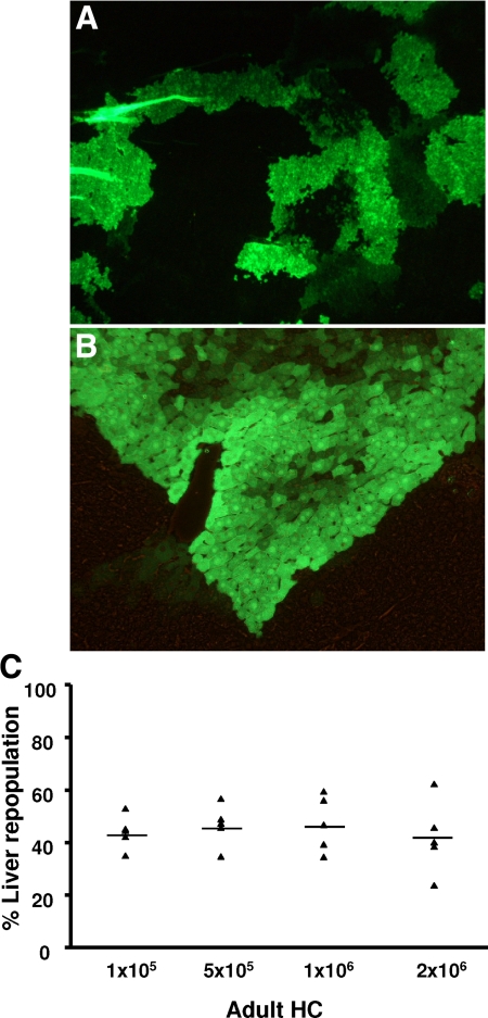
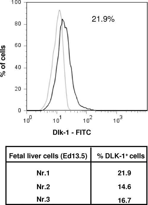
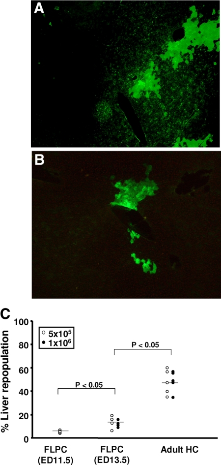
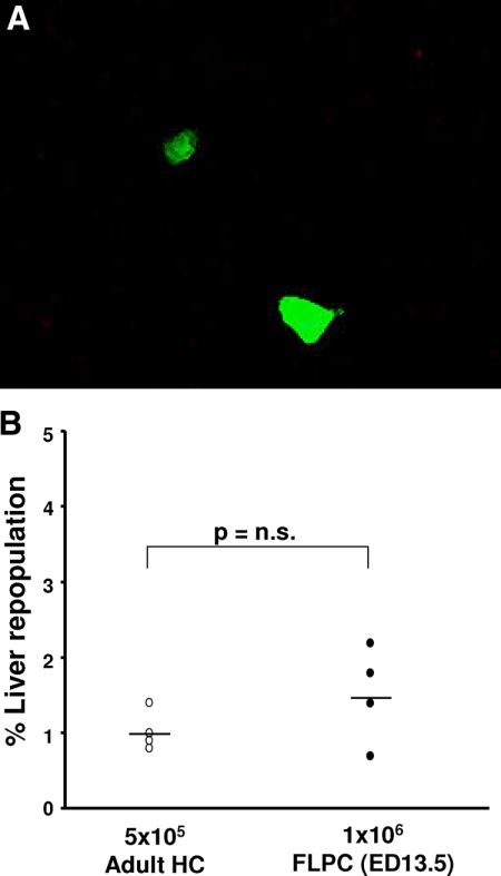
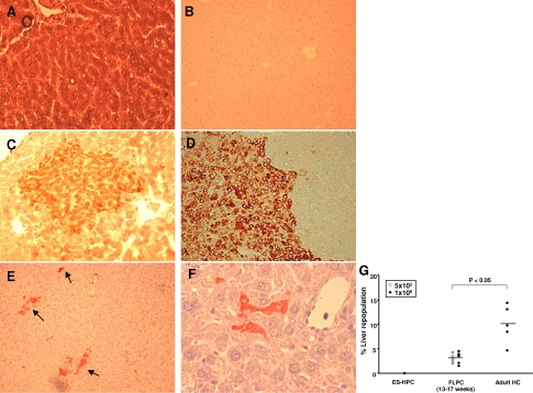
Fetal liver progenitor cell suspensions (FLPC) and hepatic precursor cells derived from embryonic stem cells (ES-HPC) represent a potential source for liver cell therapy. However, the relative capacity of these cell types to engraft and repopulate a recipient liver compared with adult hepatocytes (HC) has not been comprehensively assessed. We transplanted mouse and human HC, FLPC, and ES-HPC into a new immunodeficient mouse strain (Alb-uPA(tg(+/-))Rag2(-/-)gamma(c)(-/-) mice) and estimated the percentages of HC after 3 months. Adult mouse HC repopulated approximately half of the liver mass (46.6 +/- 8.0%, 1 x 10(6) transplanted cells), whereas mouse FLPC derived from day 13.5 and 11.5 post conception embryos generated only 12.1 +/- 3.0% and 5.1 +/- 1.1%, respectively, of the recipient liver and smaller cell clusters. Adult human HC and FLPC generated overall less liver tissue than mouse cells and repopulated 10.0 +/- 3.9% and 2.7 +/- 1.1% of the recipient livers, respectively. Mouse and human ES-HPC did not generate HC clusters in our animal model. We conclude that, in contrast to expectations, adult HC of human and mouse origin generate liver tissue more efficiently than cells derived from fetal tissue or embryonic stem cells in a highly immunodeficient Alb-uPA transgenic mouse model system. These results have important implications in the context of selecting the optimal strategy for human liver cell therapies.


Figures

Figure 1
Figure 1
Microscopic analysis of histological sections (5 μm) from a formalin-fixed liver of a recipient mouse transplanted with 5 × 10⁵ adult EGFP transgenic mouse HC (original magnification, ×200, A; ×400, B). Native EGFP fluorescence, as shown here, was confirmed in selective sections by anti-EGFP immunofluorescence (not shown). To test the relevance of transplanted cell numbers, 1 × 10⁵, 5 × 10⁵, 1 × 10⁶, and 2 × 10⁶ EGFP–transgenic mouse HC were transplanted into Alb-uPAtg(+/−)Rag2(−/−)γc(−/−) mice via intrasplenic injection. C: Three months after transplantation the number of EGFP transgenic HC detected in the recipient liver and expressed as percentage of the total number of HC was not significantly different in the four analyzed groups of animals (n = 5; p = not significant).
Figure 2
Figure 2
Murine FLPC suspensions were stained with Dlk-1 antibody and an appropriate isotype. Three representative samples were analyzed and the number of Dlk-1 positive cells were calculated.
Figure 3
Figure 3
Fluorescence microscopy of a representative formalin-fixed liver section with EGFP transgenic cell clusters from a mouse transplanted with ED 13.5 FLPC (original magnification ×200, A) and ED11.5 FLPC (original magnification ×400, B). Significantly different results were obtained between animals of groups 1 and 2, which were injected with ED 11.5 and 13.5 FLPC, respectively, as compared with animals in group 3, which were transplanted with adult HC. Results of groups 1 and 2 were not statistically different (C). The average size of the cell clusters was smaller than those observed in animals transplanted with adult mouse HC. Sorted ES-HPC did not generate sizable cell clusters in the recipient livers.
Figure 4
Figure 4
Mouse ED 13.5 FLPC and adult mouse HC were transplanted into Alb-uPAtg(−/−)Rag2(−/−)γc(−/−) mice, which do not show a liver pathology. Mostly single doublet cells were identified in the recipient livers after 3 months (original magnification ×400, A). The percentage of cells derived from FLPC and adult HC was low (<2%) and statistically not different in the two groups of animals (B).
Figure 5
Figure 5
Microscopic analysis of representative histological sections derived from human liver tissue (A), liver tissue from nontransplanted Alb-uPAtg(+/−)Rag2(−/−)γc(−/−) (B), and from livers of Alb-uPAtg(+/−)Rag2(−/−)γc(−/−) mice transplanted with human cells (CE). Human liver tissue stained uniformly positive for human albumin antigen (A). Staining was absent in nontransplanted mouse liver (B). Transplanted adult human HC formed mostly medium-sized human albumin (C, dark red colored cells) and human cytokeratin (CK) 18 (D, brown colored cells) antigen positive cell clusters surrounded by mouse liver tissue. Transplanted human FLPC (stained for human albumin, dark red color) formed single cells, which were scattered throughout the liver, and small clusters of HC (arrows) (E and F). The percentage of liver tissue repopulation in Alb-uPAtg(+/−)Rag2(−/−)γc(−/−) mice transplanted with 1 × 10⁶ human ES-HPC, 5 × 10⁵ to 1 × 10⁶ human FLPC and 1 × 10⁶ adult human HC is shown (G). The number of human albumin positive HC expressed as the average percentage of the total number of HC per microscopic field in liver sections from recipient mice was significantly different for FLPC and adult HC. Nonsorted ES-HPC did not form albumin/CK 18 positive cells in the recipient liver (not shown).
Figure 6
Figure 6
Detection of α–fetoprotein (AFP) expressing cells in human fetal liver tissue (week 17) by immunohistology. Fetal liver tissues were fixed in 4% paraformaldehyde and embedded in paraffin. Six micron thick sections were analyzed for the expression of human AFP by using a monoclonal mouse anti-human AFP (1:200) (AbD Serotec) antibody. The AFP expressing hepatoblasts (brown color) represent ∼20% of the cells in representative sections (A). Human FLPC suspensions were stained with Dlk-1 antibody and an appropriate isotype control (B). Three representative samples were analyzed and the number of Dlk-1 positive cells calculated (C).

References

    1. Gupta S, Rajvanshi P, Lee CD. Integration of transplanted hepatocytes into host liver plates demonstrated with dipeptidyl peptidase IV-deficient rats. Proc Natl Acad Sci USA. 1995;92:5860–5864. - PMC - PubMed
    1. Gupta S, Rajvanshi P, Sokhi R, Slehria S, Yam A, Kerr A, Novikoff PM. Entry and integration of transplanted hepatocytes in rat liver plates occur by disruption of hepatic sinusoidal endothelium. Hepatology. 1999;29:509–519. - PubMed
    1. Weglarz TC, Degen JL, Sandgren EP. Hepatocyte transplantation into diseased mouse liver: kinetics of parenchymal repopulation and identification of the proliferative capacity of tetraploid and octaploid hepatocytes. Am J Pathol. 2000;157:1963–1974. - PMC - PubMed
    1. Grompe M. Principles of therapeutic liver repopulation. J Inherit Metab Dis. 2009;29:421–425. - PubMed
    1. Fox IJ, Chowdhury JR, Kaufman SS, Goertzen TC, Chowdhury NR, Warkentin PI, Dorko K, Sauter BV, Strom SC. Treatment of the Crigler-Najjar syndrome type I with hepatocyte transplantation. N Engl J Med. 1998;338:1422–1426. - PubMed

Publication types

MeSH terms