. 2009 Oct;41(10):1144-9.
doi: 10.1038/ng.441. Epub 2009 Aug 30.

Tissue regenerative delays and synthetic lethality in adult mice after combined deletion of Atr and Trp53

Affiliations

Tissue regenerative delays and synthetic lethality in adult mice after combined deletion of Atr and Trp53

Yaroslava Ruzankina et al. Nat Genet. 2009 Oct.

Abstract

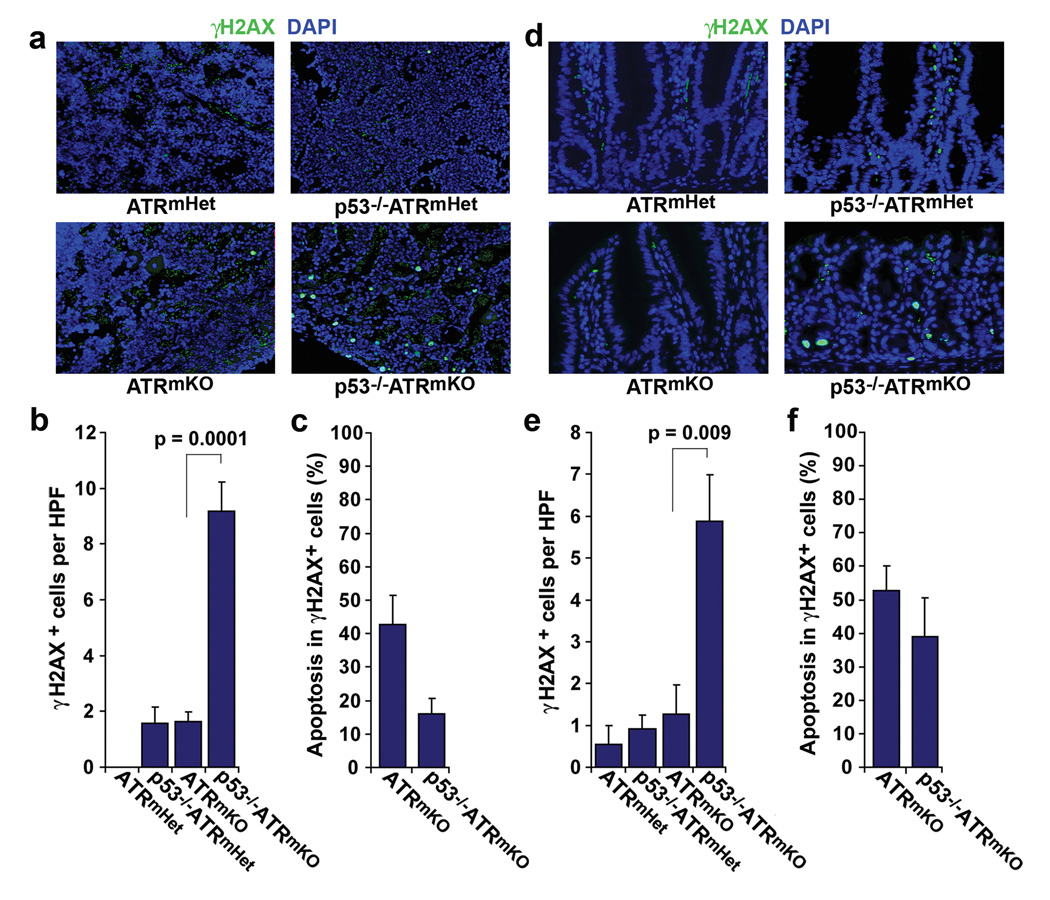
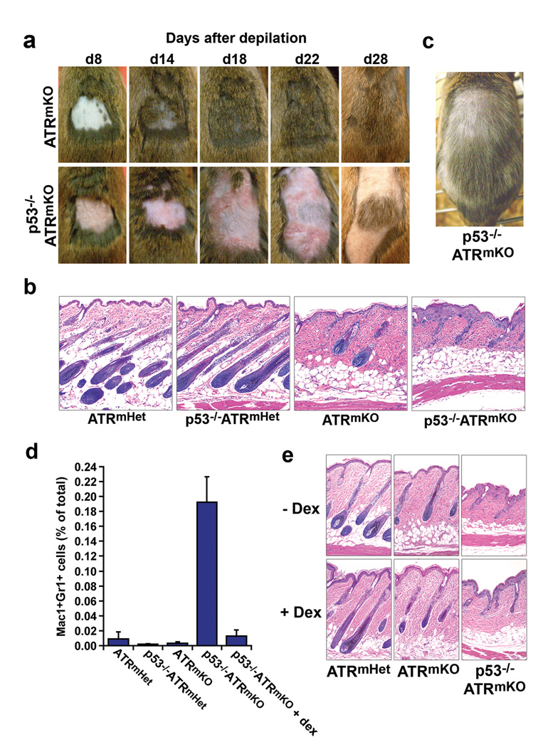
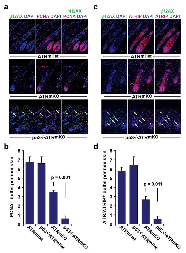
Trp53 loss of function has previously been shown to rescue tissue maintenance and developmental defects resulting from DNA damage or DNA-repair gene mutations. Here, we report that p53 deficiency severely exacerbates tissue degeneration caused by mosaic deletion of the essential genome maintenance regulator Atr. Combined loss of Atr and p53 (Trp53(-/-)Atr(mKO)) led to severe defects in hair follicle regeneration, localized inflammation (Mac1(+)Gr1(+) infiltrates), accelerated deterioration of the intestinal epithelium and synthetic lethality in adult mice. Tissue degeneration in Trp53(-/-)Atr(mKO) mice was characterized by the accumulation of cells maintaining high levels of DNA damage. Moreover, the elevated frequency of these damaged cells in both progenitor and downstream compartments in Trp53(-/-)Atr(mKO) skin coincided with delayed compensatory tissue renewal from residual ATR-expressing cells. Together, our results indicate that the combined loss of Atr and Trp53 in adult mice leads to the accumulation of highly damaged cells, which, consequently, impose a barrier to regeneration from undamaged progenitors.


Figures

Figure 1
Figure 1
Mosaic ATR deletion in adult mice is synthetic lethal with p53 deficiency. (a) Kaplan-Meier representation of survival following acute, mosaic ATR deletion in p53+/+ and p53−/− mice. Approximately 72% of ATRmKO mice survive the immediate period following TAM treatment, while less than 6% of p53−/−ATRmKO mice (1 in 17) were viable beyond this time. (b) The single surviving p53−/−ATRmKO mouse (from part a) and a comparably treated ATRmKO mouse 1 month after TAM treatment. (c) H&E stained sections of the humeral bone from mice of the indicated genotypes 6 days after TAM treatment, 200×; magnification. (d) Absolute number of myeloid cells (Gr1+) and T cells (CD3, CD4, or CD8+) obtained from 4 hindlimb bones 6 days after TAM treatment (n = 4–5 mice per genotype). The loss of myeloid cells from the bone marrow of ATRmKO and p53−/−ATRmKO relative to each respective ATRflox/+ control was significant (P = 0.01 and 0.03, respectively). (e) H&E stained sections of intestines from mice of the indicated genotypes 6 days after TAM treatment, 200× magnification. (f) Abundance of the ATRflox-recombined allele (ATRΔ) in the brain, bone marrow, and intestines 6 days after TAM treatment (n = 7–12 mice per genotype). The frequency of ATRflox recombination was determined by quantitative PCR amplification of the ATRflox allele from genomic DNA isolated from each tissue (described in Methods). P values from t tests comparing the mean frequencies of ATRΔ in p53−/−ATRmKO and ATRmKO mice were 0.047 and 0.149 in the bone marrow and intestines, respectively. Error bars in both d and f represent the S.E.M. of each data set.
Figure 2
Figure 2
Accumulation of damaged cells in the bone marrow and intestines of p53−/−ATRmKO mice. (a) Representative γH2AX (green) and DAPI (blue) stained sections of the humeral bones from mice of the indicated genotypes 6 days after TAM treatment. Autofluorescent red blood cells (green, DAPI negative) in the sinusoidal spaces of the marrow were excluded from further analysis. (b) Quantification of γH2AX-positive cells in the bone marrow. The abundance of γH2AX-positive cells was determined from 6–10 high power field images (HPF, 200×) of indicated mice (n = 3–6 mice per genotype). Only nucleated (DAPI-positive) cells were scored in this analysis. (c) Frequency of γH2AX-positive cells staining positive for cleaved caspase-3, as determined by co-immunostaining (also see Supplementary Fig. 3a). (d) Representative γH2AX (green) and DAPI (blue) stained intestinal sections from mice of the indicated genotypes 6 days after TAM treatment. Autofluorescent red blood cells (green, DAPI negative) in the vasculature and stroma were excluded from quantifications. (e) Quantification of γH2AX-positive cells in intestinal epithelium (n = 2–6 mice per genotype). The abundance of γH2AX-positive cells was determined as in b. (f) Frequency of γH2AX-positive cells staining positive for cleaved caspase-3, as determined by co-immunostaining (also see Supplementary Fig. 3b). Error bars in b, c, e and f represent the S.E.M. of each data set.
Figure 3
Figure 3
p53 deficiency dramatically delays hair follicle regeneration following localized ATR deletion in the skin and leads to acute inflammation. (a) Time course following depilation of 4-OHT-treated skins of ATRflox/− and p53−/−ATRflox/− mice. An appreciable delay in hair regeneration after depilation was observed in p53−/−ATRmKO skin and was accompanied by the outward manifestations of inflammation (redness, swelling, exfoliation). These phenotypes were consistently observed in p53−/−ATRmKO mice (n = 6–12 mice per genotype). (b) Representative H&E stained sections of the epidermis 8 days after depilation. The number and quality of regenerating follicles were compromised in the p53−/−ATRmKO skin relative to ATRmKO or ATRflox/+ control skins (n = 5–7 mice per genotype). (c) Regenerated hair in p53−/−ATRmKO skin 48 days after depilation. Appreciable hair graying was observed in p53−/−ATRmKO skin in the depilated and surrounding 4-OHT treatment regions (n = 6 mice). (d) Quantification of Gr1+Mac1+ inflammatory cells in the epidermis 8 days after depilation (n = 4–5 mice per genotype). Total epidermal cells were isolated (Methods), stained with antibodies to Gr1 and Mac1 (CD11b), and the frequency of Gr1+Mac1+ cells was quantified by flow cytometry. Gr1+Mac1+ cells accumulated significantly in p53−/−ATRmKO skin (P = 3 × 10⁻⁷), and dexamethasone pretreatment of p53−/−ATRmKO skin (+ dex) prevented Gr1+Mac1+ cell accumulation, P = 0.003. Error bars, s.e.m. (e) H&E stained sections of skin 8 days after depilation in the presence or absence of dex. Mice treated with dex or left untreated were sacrificed 8 days after depilation. Delayed follicle regeneration in p53−/−ATRmKO skin was unaffected by dexamethasone treatment (n = 4 mice per genotype).
Figure 4
Figure 4
p53−/−ATRmKO skin is characterized by the persistent accumulation of γH2AX-positive cells. (a) Frequency of the ATRflox-recombined allele (ATRΔ) in epidermal isolates at different time points after depilation (n = 3–13 mice per genotype per time point). Recombination of the ATRflox allele was quantified by qPCR amplification of genomic DNA isolated from total epidermal cells (described in Methods). (b) Frequency of γH2AX-positive epidermal stem and progenitor cells (CD34+α-6-integrin+) isolated from skin 4 days and 8 days following depilation. The difference in mean abundance of γH2AX-positive cells between p53−/−ATRmKO and ATRmKO skins 8 days post-depilation was significant (P = 0.023). (c) Representative γH2AX (brown) and hematoxylin (blue) stained sections of the skin 4 days and 8 days after depilation. (d) Quantification of γH2AX-positive cells in the hair follicles and epidermis of depilated skin. Frequency of γH2AX-positive cells was determined in 6–10 sections per mouse (n=3–5 mice per genotype for each time point). Note the elevated frequency of γH2AX-positive cells in both ATRmKO and p53−/−ATRmKO skins 4 days post-depilation; however, this significant increase was maintained only in p53−/−ATRmKO skin 4 days later (day 8 post-depilation). The difference in mean abundance of γH2AX-positive cells between p53−/−ATRmKO and ATRmKO skins 8 days post-depilation was significant (P = 0.013). Increased frequency of γH2AX-positive cells in p53−/−ATRmKO skin was observed both in the presence and absence of dexamethasone (Supplementary Fig. 4).
Figure 5
Figure 5
p53 is required for efficient compensatory renewal following ATR deletion. (a) Representative PCNA (red), γH2AX (green), and DAPI (blue) stained sections of skin 8 days after depilation. γH2AX-positive cells are indicated (white arrows). (b) Quantification of regenerating PCNA-positive bulbs per mm skin. (c) Follicle regeneration in ATRmKO skin by ATR/ATRIP-expressing cells. Representative ATRIP (red), γH2AX (green), and DAPI (blue) stained sections of skin 8 days after depilation. ATR and ATRIP form an interdependent complex in which ATR is required for ATRIP stability. γH2AX-positive cells are indicated (white arrows). (d) Quantification of ATR/ATRIP-expressing bulbs per mm skin. Analyses and quantifications for b and d were performed on 6–12 sections per mouse (n=3 mice per genotype). Error bars represent S.E.M. for each data set.

References

    1. Wlodarski P, et al. Role of p53 in hematopoietic recovery after cytotoxic treatment. Blood. 1998;91:2998–3006. - PubMed
    1. Bender CF, et al. Cancer predisposition and hematopoietic failure in Rad50(S/S) mice. Genes Dev. 2002;16:2237–2251. - PMC - PubMed
    1. Chin L, et al. p53 deficiency rescues the adverse effects of telomere loss and cooperates with telomere dysfunction to accelerate carcinogenesis. Cell. 1999;97:527–538. - PubMed
    1. Frank KM, et al. DNA ligase IV deficiency in mice leads to defective neurogenesis and embryonic lethality via the p53 pathway. Mol Cell. 2000;5:993–1002. - PubMed
    1. Gao Y, et al. Interplay of p53 and DNA-repair protein XRCC4 in tumorigenesis, genomic stability and development. Nature. 2000;404:897–900. - PubMed

Publication types

MeSH terms

Substances