Comparative Study
. 2009 Sep;158(1):300-13.
doi: 10.1111/j.1476-5381.2009.00328.x.

Cross-regulation between beta 1- and beta 3-adrenoceptors following chronic beta-adrenergic stimulation in neonatal rat cardiomyocytes

Affiliations
Comparative Study

Cross-regulation between beta 1- and beta 3-adrenoceptors following chronic beta-adrenergic stimulation in neonatal rat cardiomyocytes

Christoph Ufer et al. Br J Pharmacol. 2009 Sep.

Abstract

Background and purpose: We have previously shown that beta-adrenoceptors continuously stimulated with noradrenaline induces an increase in beta(3)-adrenoceptors (G alpha(i)PCRs) and a decrease in beta(1)-adrenoceptors (G alpha(s)PCRs) at functional, genomic and protein levels. This compensatory modification induced by noradrenaline is probably one of the consequences of cardiac depression observed in heart disease. Therefore, we investigated further the interaction between beta(1)- and beta(3)-adrenoceptors in neonatal rat cardiomyocytes.

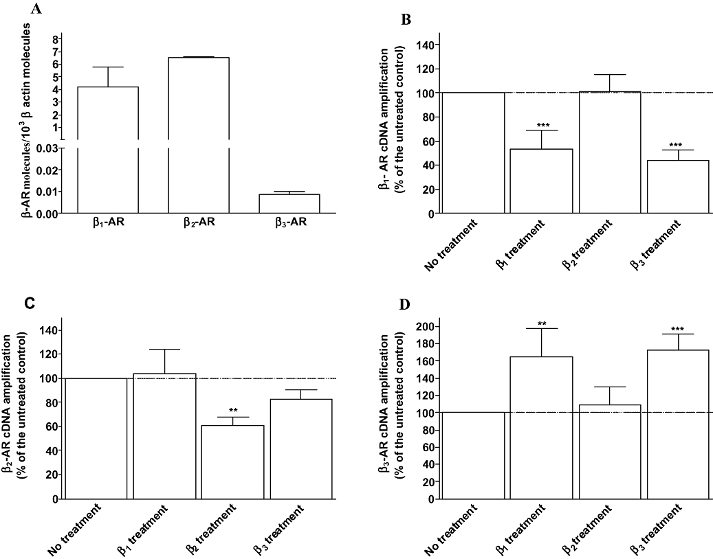
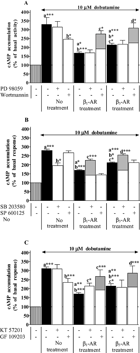
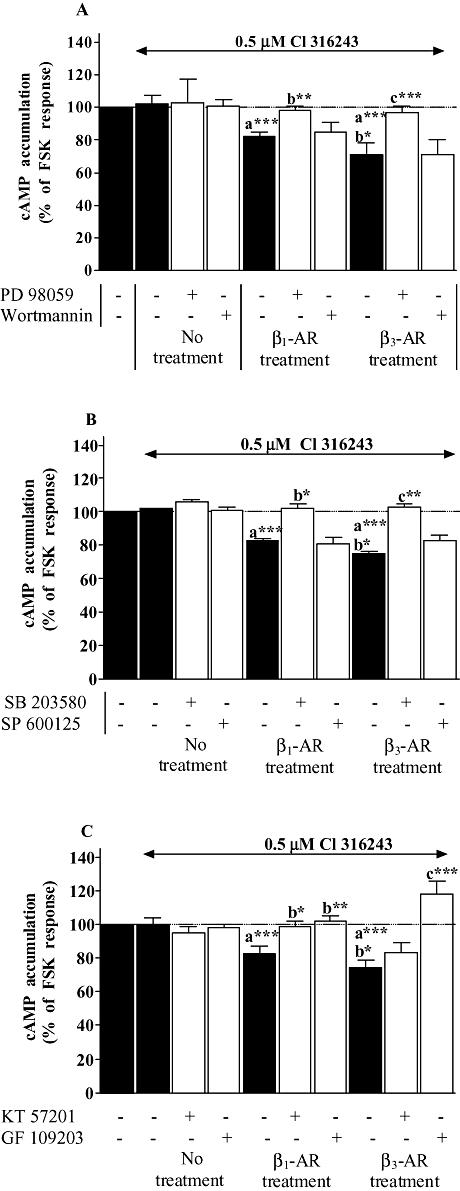
Experimental approach: Functional studies were performed by cyclic adenosine monophosphate (cAMP) accumulation assays in cells untreated or treated with dobutamine and ICI 118551 (beta(1)-adrenoceptor) or CL-3162436243 (beta(3)-adrenoceptor) for 24 h in the presence or absence of protein kinase inhibitors. Beta-adrenoceptor and protein kinase expression was monitored by quantitative reverse transcription-polymerase chain reaction (RT-PCR) and by Western blotting, respectively.

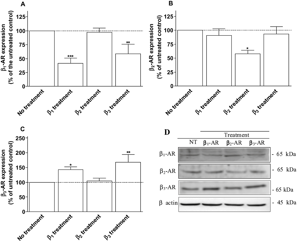
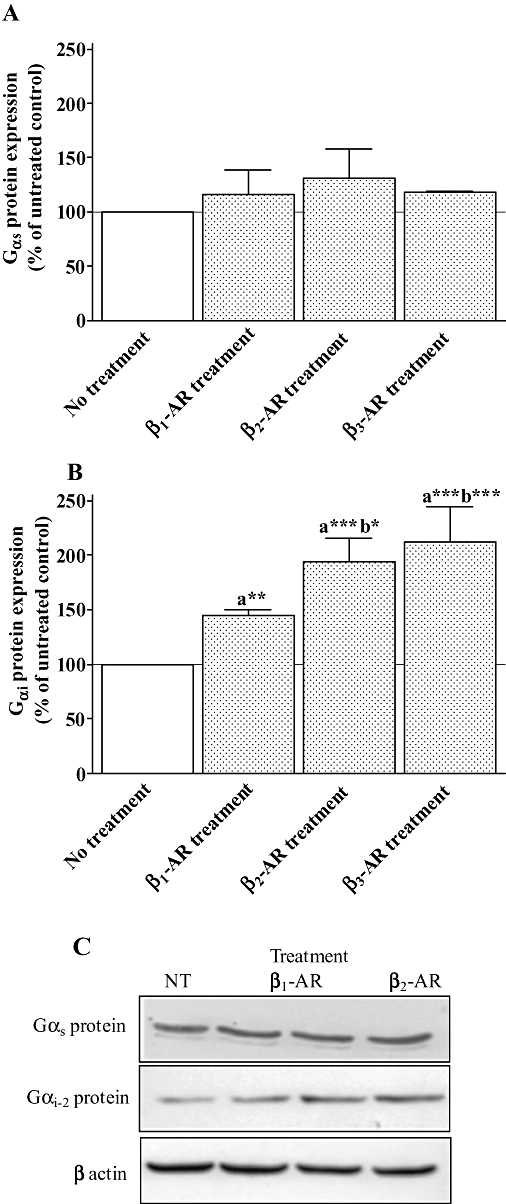
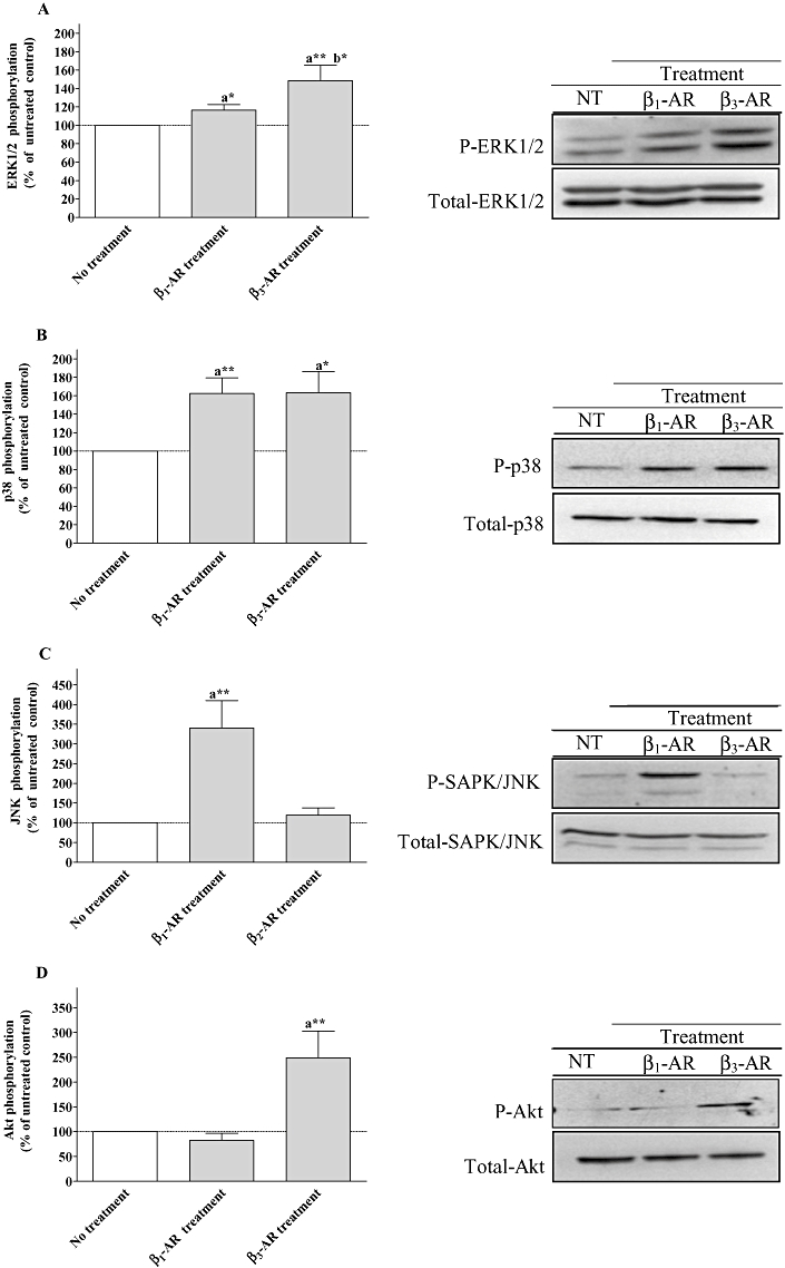
Key results: Chronic beta(1)- or beta(3)-adrenoceptor stimulation reduced beta(1)-adrenoceptor-mediated cAMP accumulation in association with a decrease in beta(1)-adrenoceptor mRNA and protein levels through protein kinase C (PKC), phosphoinositide 3-kinase (PI3K) and p38 mitogen-activated protein kinase (p38MAPK) activation. In contrast, both treatments induced an increase in beta(3)-adrenoceptor expression and beta(3)-adrenoceptor-inhibited forskolin response through PKC, extracellular-signal-regulated kinases 1 and 2 (ERK1/2) and p38MAPK phosphorylation, although no beta(3)-adrenoceptor response was observed in untreated cells. ERK1/2 and p38MAPK were activated by both treatments. The modulation of beta(1)- or beta(3)-adrenoceptor function did not require stress-activated protein kinase/c-Jun N-terminal kinase (SAPK/JNK) although chronic beta(1)-adrenoceptor stimulation activated SAPK/JNK. Beta(3)-adrenoceptor treatment activated Akt although PI3K was not involved in beta(3)-adrenoceptor up-regulation.

Conclusion and implications: We show for the first time that chronic beta(1)- or beta(3)-adrenoceptor stimulation leads to the modulation of beta(1)- and beta(3)-adrenoceptors by a cross-regulation involving PKC, PI3K p38MAPK and MEK/ERK1/2 pathway, and through protein kinase A when beta(1)-adrenoceptors are chronically activated.


Figures

Figure 1
Figure 1
Activation of cAMP accumulation induced by dobutamine, β1-selective agonist (A) and procaterol, β2-selective agonist (B), and effect of the selective β3-adrenoceptor agonist CL-316243 on forskolin (Forsk)-induced cAMP accumulation (C) in neonatal rat cardiomyocytes untreated, treated with 10 µM dobutamine in the presence of 1 µM ICI 118551, 10 µM procaterol in the presence of 1 µM CGP 20712A or 2 µM CL-316243 for 24 h. cAMP accumulation was measured as described under Methods. Data are expressed as percentage of the basal level of cAMP accumulation (100%, A, B) or as the percentage of 1.5 µM forskolin response in the absence of agonist (100%, C). Each point represents the mean ± SEM of four to six experiments performed in duplicate.
Figure 3
Figure 3
Expression of β1- (A), β2- (B) and β3-adrenoceptors (C) obtained from neonatal rat cardiomyocytes untreated (control), treated with 10 µM dobutamine in the presence of 1 µM ICI 118551 (β1 treatment), 10 µM procaterol in the presence of 1 µM CGP 20712A (β2 treatment) or 2 µM CL-316243 (β3 treatment) for 24 h. Cell membrane homogenates were monitored by Western blotting for β1-, β2- and β3-adrenoceptors as described under Methods. The same samples were also analysed on separate blots using an antibody that recognizes β actin to confirm equal loading on each lane. Representative immunoblots for each proteins are shown in panel C. Data are expressed as the percentage of untreated cardiomyocytes (100%) following the calculation of β-adrenoceptor/β-actin ratio for each lane. The combined results (panel A, B and C) obtained from densitometric analysis of blots represent the mean ± SEM of five to seven independent experiments. *P < 0.05 and **P < 0.01 versus untreated control.
Figure 2
Figure 2
Quantitative expression of β-adrenoceptors in untreated neonatal rat cardiomyocytes (A) and expression of β1- (B), β2- (C) and β3-adrenoceptors (D) mRNA obtained from untreated (control), treated with 10 µM dobutamine in the presence of 1 µM ICI 118551 (β1 treatment), 10 µM procaterol in the presence of 1 µM CGP 20712A (β2 treatment) or 2 µM CL-316243 (β3 treatment) for 24 h. Total RNA was prepared, reverse transcription and real time PCR were carried out as described under Methods. Values were expressed as β-adrenoceptor molecules × 10−3β actin molecules in A and normalized to β-actin molecules and expressed as the percentage of untreated cardiomyocytes (100%) in B, C and D. Each point represents the mean ± SEM of five to six independent experiments. **P < 0.01 and ***P < 0.001 versus untreated control.
Figure 4
Figure 4
Expression of Gαs and Gαi proteins obtained from neonatal rat cardiomyocytes untreated (control), treated with 10 µM dobutamine in the presence of 1 µM ICI 118551 (β1 treatment), 10 µM procaterol in the presence of 1 µM CGP 20712A (β2 treatment) or 2 µM CL-316243 (β3 treatment) for 24 h. Cell membrane homogenates were monitored by Western blotting for Gαs (A) and Gαi (B) proteins as described under Methods. The same samples were also analysed on separate blots using an antibody that recognizes β actin to confirm equal loading on each lane. Representative immunoblots for each proteins are shown in panel C. Data are expressed as the percentage of untreated cardiomyocytes (100%) following the calculation of β-adrenoceptor/β-actin ratio for each lane. The combined results (panels A and B) obtained from densitometric analysis of blots represent the mean ± SEM of four to six independent experiments. *P < 0.05, **P < 0.01 and ***P < 0.001, (a) versus untreated control and (b) versus β1-adrenoceptor treatment.
Figure 5
Figure 5
Effect of kinase inhibitors on dobutamine-induced cAMP accumulation in neonatal rat cardiomyocytes untreated, treated with 10 µM dobutamine in the presence of 1 µM ICI 118551 (β1 treatment), or 2 µM CL-316243 (β3 treatment) for 24 h. In addition, untreated and treated cells were incubated in panel A with 50 µM PD 98059 (MEK inhibitor) or 100 nM wortmannin (phosphoinositide 3-kinase inhibitor), in panel B with 10 µM SB 203580 (p38 MAPK inhibitor) or 10 µM SP 600125 (JNK inhibtor) and in panel C with 1 µM KT 57201 (PKA inhibitor) or 10 µM GF 109203 (PKC inhibitor). cAMP accumulation was measured as described under Methods. Data are expressed as the percentage of the basal level of cAMP accumulation (set at 100%, striped bars). Black and open bars represent cAMP accumulation induced by dobutamine in the absence and presence of protein kinase inhibitors in all groups respectively. Wortmanin (A), SB 203580 (B) and GF 109203 (C) elicited a significant decrease in cAMP accumulation induced by dobutamine in untreated cardiomyocytes. Grey filled bars represent the data corrected from the effect of these inhibitors on dobutamine response in untreated cardiomyocytes in order to consider only the effect of β1- or β3-adrenoceptor treatments on the modulation of β1-adrenoceptor response (decrease in untreated cells + value of treated cardiomyocytes for each individual experiments). Each bar represents the mean ± SEM of six to seven independent experiments. *P < 0.05, **P < 0.01 and ***P < 0.001, (a) versus basal level, (b) versus dobutamine response in untreated cells, (c) versus dobutamine response in β1-adrenoceptor-treated cells and (d) versus dobutamine response in β3-adrenoceptor-treated cells.
Figure 6
Figure 6
Effect of kinase inhibitors on CL-3162436243-inhibited forskolin response in neonatal rat cardiomyocytes untreated, treated with 10 µM dobutamine in the presence of 1 µM ICI 118551 (β1 treatment), or 2 µM CL-316243 (β3 treatment) for 24 h. In addition, untreated and treated cells were incubated in panel A with 50 µM PD 98059 (MEK inhibitor) or 100 nM wortmannin (phosphoinositide 3-kinase inhibitor), in panel B with 10 µM SB 203580 (p38 MAPK inhibitor) or 10 µM SP 600125 (JNK inhibtor) and in panel C with 1 µM KT 57201 (PKA inhibitor) or 10 µM GF 109203 (PKC inhibitor). cAMP accumulation was measured as described under Methods. Data are expressed as the percentage of 1.5 µM forskolin (FSK) response in the absence of agonist (100%, striped bars). Black and open bars represent the inhibition of FSK-mediated cAMP accumulation induced by CL-316243 in the absence and presence of protein kinase inhibitors in all groups respectively. Each bar represents the mean ± SEM of six to seven independent experiments. *P < 0.05, **P < 0.01 and ***P < 0.001, (a) versus CL-316243 response in untreated cells, (b) versus CL-316243 response in β1-adrenoceptor treated cells and (c) versus CL-316243 response in β3-adrenoceptor treated cells.
Figure 7
Figure 7
Effect of β1-adrenoceptor and β3-adrenoceptor treatments on ERK1/2, p38 MAPK, stress-activated protein kinase/c-Jun N-terminal kinase (SAPK/JNK) and Akt activation in neonatal rat cardiomyocytes. The cells were untreated or treated with 10 µM dobutamine in the presence of 1 µM ICI 118551 (β1-AR treatment), or 2 µM CL-316243 (β3-AR treatment) for 24 h. Following the treatments, cell lysates were analysed by Western blotting as described under Methods using phospho-specific antibodies. The same samples were also analysed on separate blot using an antibody that recognizes both unphosphorylated (total) and phosphorylated kinases to confirm equal loading on each lane. Data are expressed as percentage of the untreated control following the calculation of the phosphorylated/total ratio for each lane. The combined results (panel A for ERK1/2, panel B for p38 mitogen-activated protein kinase, panel C for SAPK/JNK and panel D for Akt) obtained from densitometric analysis of blots, represent the mean ± SEM of for to six independent experiments. *P < 0.05 and **P < 0.01, (a) versus untreated control, (b) versus β1-adrenoceptor treated cells.

References

    1. Alexander SPH, Mathie A, Peters JA. Guide to Receptors and Channels (GRAC), 3rd edition (2008 revision) Br J Pharmacol. 2008;153(Suppl. 2):S1–S209. - PMC - PubMed
    1. Barbier J, Rannou-Bekono F, Marchais J, Tanguy S, Carre F. Alteration of beta(3)-adrenoceptors expression and their myocardial functional effects in physiological model of chronic exercise-induced cardiac hypertrophy. Mol Cell Biochem. 2007;300:69–75. - PubMed
    1. Bensaid M, Kaghad M, Rodriguez M, Le Fur G, Caput D. The rat beta 3-adrenergic receptor gene contains an intron. FEBS Lett. 1993;318:223–226. - PubMed
    1. Cheng H-J, Zhang Z-H, Onishi K, Ukai T, Sane DC, Cheng C-P. Upregulation of functional β3-adrenergic receptor in the failing canine myocardium. Circ Res. 2001;89:599–606. - PubMed
    1. Cohn JN, Levine TB, Olivari MT, Garberg V, Lura D, Francis GS, et al. Plasma norepinephrine as a guide to prognosis in patients with chronic congestive heart failure. N Engl J Med. 1984;311:819–823. - PubMed

Publication types

MeSH terms

Substances