. 2009 Oct;126(10):791-803.
doi: 10.1016/j.mod.2009.08.003. Epub 2009 Aug 29.

Wnt signaling maintains the notochord fate for progenitor cells and supports the posterior extension of the notochord

Affiliations

Wnt signaling maintains the notochord fate for progenitor cells and supports the posterior extension of the notochord

Kanako Ukita et al. Mech Dev. 2009 Oct.

Abstract

The notochord develops from notochord progenitor cells (NPCs) and functions as a major signaling center to regulate trunk and tail development. NPCs are initially specified in the node by Wnt and Nodal signals at the gastrula stage. However, the underlying mechanism that maintains the NPCs throughout embryogenesis to contribute to the posterior extension of the notochord remains unclear. Here, we demonstrate that Wnt signaling in the NPCs is essential for posterior extension of the notochord. Genetic labeling revealed that the Noto-expressing cells in the ventral node contribute the NPCs that reside in the tail bud. Robust Wnt signaling in the NPCs was observed during posterior notochord extension. Genetic attenuation of the Wnt signal via notochord-specific beta-catenin gene ablation resulted in posterior truncation of the notochord. In the NPCs of such mutant embryos, the expression of notochord-specific genes was down-regulated, and an endodermal marker, E-cadherin, was observed. No significant alteration of cell proliferation or apoptosis of the NPCs was detected. Taken together, our data indicate that the NPCs are derived from Noto-positive node cells, and are not fully committed to a notochordal fate. Sustained Wnt signaling is required to maintain the NPCs' notochordal fate.


Figures

Figure 1
Figure 1. Proliferation of NPCs during development
(A, B) BrdU-labeling of (A) the E7.75 node (indicated by a dotted line) and (B) the E10.5 tail region of normal embryos. The posterior end of the notochord is surrounded by a dotted line. A representative E9.25 tail region is shown in Figure 5I. (C) Ratios of BrdU-positive cells in the node (E7.75) or at the posterior end of the notochord (E9.25 and E10.5). Scale bar, 100 μm for A and B. Abbreviations: hg, hindgut; n, node; nc, notochord; nt, neural tube; ps, primitive streak.
Figure 2
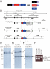
Figure 2. Production of the NotonmCherry-CreERT2/+ mouse
(A) Production of three proteins from a single transcript. 2A-peptide-containing proteins are self-cleaved downstream of the 2A peptide. Therefore, the modified Noto-locus, which has a single coding sequence for a Noto-2A-nuclear mCherry-2A-CreERT2 protein, will produce three proteins: Noto-2A, nuclear mCherry-2A, and CreERT2. (B) Targeting strategy. The NotonmCherry-CreERT2 allele was generated by homologous recombination in ES cells. Exons are indicated by filled and open boxes, and coding regions by filled boxes. Green triangles represent loxP sites. 5′ and 3′ indicate the approximate positions of probes for Southern hybridization. Abbreviations: H, Hind III; mC, ERAV 2A peptide followed by nuclear mCherry; CreER, TaV 2A peptide followed by CreERT2; ACE-Cre, a cassette carrying the testis-specific ACE promoter-Cre-poly A signal; neo, an expression cassette for the neomycin-resistance gene; DT-A, MC1-DT-A-poly signal cassette. The ACN cassette consists of tACE-Cre and neo cassettes flanked by loxP sites, and this cassette is removed in the mouse testis during germline transmission. Arrowheads indicate the approximate positions of PCR primers. F2, SVloxP-3′-F2; R2, Noto-PC-3′-R2; nF, Noto genotype F; eF, ERT2 genotype F; nR, Noto genotype R. (C-E) Southern blot analysis of homologous recombination in ES cells. Hind III-digested ES genomes were separated on a 0.3% agarose gel and blotted to nylon membranes. Hybridization with the 5′ probe (C) and 3′ probe (D) gave two bands that corresponded to the predicted lengths of the wild-type (wt) and knock-in (ki) alleles. Hybridization with the internal (neo) probe (E) gave a single band corresponding to the knock-in allele. (F) Genotype determination of NotonmCherry-CreERT2 mice by PCR. Note that the ACN cassette was removed in knock-in mice #1 and #4, while knock-in ES cells produced only a wild-type band, because the ACN cassette prevented PCR amplification of the mutant allele.
Figure 3
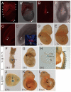
Figure 3. Genetic lineage tracing of Noto-expressing cells
(A-C) Expression of nuclear mCherry in NotonmCherry-CreERT2/+ embryos. The mCherry signal was observed in the nuclei of node cells and in the posterior head process (arrowheads) at E7.5 (A, A’). Stars indicate auto-fluorescence in the visceral endoderm, which was not derived from mCherry. The mCherry signal was observed in the node (arrowhead) of E8.5 embryos (B, B’) and the posterior notochord-like tissue in the tail bud (arrowheads) at E9.5 (C, C’) embryos. The inset in C’ shows the immunofluorescence staining of mCherry protein (red) in a cross-section of E9.5 tail. Nuclei were counterstained with DAPI (blue). (D, E) Cre activity depends on Tx administration in NotonmCherry-CreERT2/+;ROSA26lacZ/+ embryos. Tx administration at E8.5 induced Cre-mediated recombination in the posterior notochord by E9.5 (D), while no recombination was observed without Tx (E). (F-I) Lineage tracing of Noto-positive cells by Tx administration at E7.75. β-galactosidase activity at E8.5 (F, ventral view), E9.5 (G, side view; H, a cross section near the posterior end of the notochord) and E13.5 (I, tail region). (J-L) Lineage tracing of Noto-positive cells by Tx administration at E6.5. β-galactosidase activity at E7.5 (J, ventral view, anterior is to the left) and E10.5 (K, L) embryos. Arrows in G, I, K and L indicate the posterior end of the notochord. Stars in G, I, and L indicate non-notochordal cells labeled with β-galactosidase. Abbreviations: a, allantois; fl, fore limb; hg, hindgut; mb, midbrain; n, node; nc, notochord; np, neural plate; nt, neural tube.
Figure 4
Figure 4. Activation of Wnt signaling in the NPCs
(A-E) Whole-mount β-galactosidase staining of TOPGAL transgenic embryos at (A) E8.5, (B) E8.75, (C) E11.5, (D) E12.5, and (E) E13.5. Arrows indicate the node or the posterior end of the notochord. (F) Sagittal section through the tail of an E9.5 TOPGAL embryo. (G-H) Whole-mount β-galactosidase staining of a ins-TOPGAL transgenic embryo at E9.5 and (H, I) sagittal sections through the tails of similar embryos. The color reaction was allowed to proceed for (G) 4 hours, (H) 45 minutes, or (I) 15 minutes. Abbreviations: en, endoderm; h, heart; n, node; nc, notochord; nt, neural tube.
Figure 5
Figure 5. Attenuation of Wnt signaling by notochord-specific ablation of the β-catenin gene
(A-D) Whole-mount β-galactosidase staining of Not-Cre;ROSA26 embryos at (A) E8.25, (B) E8.5, and (C) E9.5. (D) Sagittal section through an E9.5 tail. (E, F) Immunofluorescent staining of β-catenin protein in (E) control and (F) Not-Cre;β-cateninflox/flox (N-Cre;β-catenin) embryos. Arrows indicate the notochord, showing the specific ablation of β-catenin. (G-J) Whole-mount β-galactosidase staining and tail-region cross-sections of E9.5 (G, I) TOPGAL and (H, J) Not-Cre;β-cateninflox/flox; TOPGAL embryos. Abbreviations: en, endoderm; fl, forelimb bud; h, heart; hg, hindgut; n, node; nc, notochord; nt, neural tube; np, neural plate.
Figure 6
Figure 6. Not-Cre;β-cateninflox/flox mutants lack a posterior notochord
(A-D) Hematoxylin and eosin staining of cross-sections through the tail regions of (A, C) control and (B, D) Not-Cre;β-cateninflox/flox (N-Cre;β-catenin) embryos at (A, B) E9.5 and (C, D) E10.5. External morphologies of E10.5 (E) control and (F) Not-Cre;β-cateninflox/flox embryos, showing the curled and shorter tail of the mutant (arrowheads). (G, H) Skeletal specimen of (G) control and (H) Not-Cre;β-cateninflox/flox neonates (P0). The interparietal bone of the posterior skull of the control neonate was lost during the staining procedure. Scale bars: 50 μm for A, B, C, and D; 2 mm for E, F; 1 cm for G, H. Abbreviations: fl, forelimb bud; hg, hindgut; hl, hindlimb bud; nc, notochord; nt, neural tube; s, somite; S1, first sacral vertebra.
Figure 7
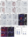
Figure 7. Not-Cre;β-cateninflox/flox embryos showed altered gene expression and unaltered cell proliferation and survival at the end of the posterior notochord
(A-V) Arrowheads indicate the posterior end of the notochord, and arrows indicate the notochord/notochord-like tissue, throughout the figure. (A-D) Whole-mount in situ hybridization of Noto and the corresponding cross-sections through the posterior region in E9.5 (A, C) control and (B, D) Not-Cre;β-cateninflox/flox (N-Cre;β-catn) embryos. (E-J) Whole-mount in situ hybridization of Brachyury and corresponding cross-sections in E9.25 (E, G, I) control and (F, H, J) Not-Cre;β-cateninflox/flox embryos. (G, H) Cross-sections through the trunk. (I, J) Cross-sections through the posterior region. (K-N) Cross-sections through the posterior region of E9.25 (K, M) control and (L, N) Not-Cre;β-cateninflox/flox embryos stained for (K, L) Shh or (M, N) Foxa1 RNA by whole-mount in situ hybridization. (O, P) Immunohistochemical staining of Foxa2 on cross-sections through the posterior region of E9.5 (O) control and (P) Not-Cre;β-cateninflox/flox embryos. (Q-R’) Immunofluorescent staining of E-cadherin on cross-sections through the posterior region of E9.25 (Q, Q’) control and (R, R’) Not-Cre;β-cateninflox/flox embryos. Q, R: merged images with DAPI (blue), Q’, R’: E-cadherin only. (S-V) (S, T) BrdU labeling and (U, V) TUNEL of the tail region of E9.25 (S, U) control and (T, V) Not-Cre;β-cateninflox/flox embryos. (W, X) Percentage of posterior-end notochord cells that were (W) BrdU-positive or (X) TUNEL-positive cells. Error bars indicate standard deviation. Scale bars: 1 mm for A, B, E, and F; 50 μm for G-R; 100 μm for S-V. Abbreviations: h, heart; hg, hindgut; hl, hindlimb bud; mg, midgut; np, neural plate; nt, neural tube.

Similar articles

Cited by

References

    1. Abdelkhalek HB, Beckers A, Schuster-Gossler K, Pavlova MN, Burkhardt H, Lickert H, Rossant J, Reinhardt R, Schalkwyk LC, Muller I, Herrmann BG, Ceolin M, Rivera-Pomar R, Gossler A. The mouse homeobox gene Not is required for caudal notochord development and affected by the truncate mutation. Genes Dev. 2004;18:1725–36. - PMC - PubMed
    1. Agathon A, Thisse C, Thisse B. The molecular nature of the zebrafish tail organizer. Nature. 2003;424:448–52. - PubMed
    1. Ang SL, Rossant J. HNF-3 β is essential for node and notochord formation in mouse development. Cell. 1994;78:561–574. - PubMed
    1. Beck CW, Slack JM. A developmental pathway controlling outgrowth of the Xenopus tail bud. Development. 1999;126:1611–20. - PubMed
    1. Beck CW, Whitman M, Slack JM. The role of BMP signaling in outgrowth and patterning of the Xenopus tail bud. Dev Biol. 2001;238:303–14. - PubMed

Publication types