. 2009 Sep;144(1-2):44-57.
doi: 10.1016/j.virusres.2009.03.020. Epub 2009 Apr 7.

Conserved structure/function of the orthoreovirus major core proteins

Affiliations

Conserved structure/function of the orthoreovirus major core proteins

Wanhong Xu et al. Virus Res. 2009 Sep.

Abstract

Orthoreoviruses are infectious agents with genomes of 10 segments of double-stranded RNA. Detailed molecular information is available for all 10 segments of several mammalian orthoreoviruses, and for most segments of several avian orthoreoviruses (ARV). We, and others, have reported sequences of the L2, all S-class, and all M-class genome segments of two different avian reoviruses, strains ARV138 and ARV176. We here determined L1 and L3 genome segment nucleotide sequences for both strains to complete full genome characterization of this orthoreovirus subgroup. ARV L1 segments were 3958 nucleotides long and encode lambda A major core shell proteins of 1293 residues. L3 segments were 3907 nucleotides long and encode lambda C core turret proteins of 1285 residues. These newly determined ARV segments were aligned with all currently available homologous mammalian reovirus (MRV) and aquareovirus (AqRV) genome segments. Identical and conserved amino acid residues amongst these diverse groups were mapped into known mammalian reovirus lambda 1 core shell and lambda 2 core turret proteins to predict conserved structure/function domains. Most identical and conserved residues were located near predicted catalytic domains in the lambda-class guanylyltransferase, and forming patches that traverse the lambda-class core shell, which may contribute to the unusual RNA transcription processes in this group of viruses.


Conflict of interest statement

The authors declare no competing interests.

Figures
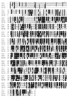
Fig. 1
Fig. 1
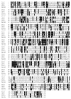
Alignment of the deduced ARV138 and ARV176 λA amino acid sequences. All 26 currently available homologous ARV λA, MRV λ1, and AqRV VP3 proteins (determined for each clone shown in Table 1) were aligned, both by T-Coffee (Notredame et al., 2000) (data not shown) and by Clustal-W (Thompson et al., 1994), with only minor differences in the alignments created by different gap penalties (data not shown). Only the two most-distant ARV, MRV, and AqRV sequences (see text for details) are shown for clarity. Clones are: MRV–T1L (GenBank no. NC_004255) and T2J (GenBank no. NC_004256); ARV–ARV138 (GenBank no. EU707933) and ARV176 (GenBank no. EU707934); AqRV–Grass Carp reovirus (GCRV) (GenBank no. AF260513) and Chum Salmon reovirus (CSRV) (GenBank No. NC_007584). Amino acid residues that are identical in at least four of the sequences are indicated by black background shading. The single letter amino acid code is used. Six previously identified putative helicase domains (labeled I–VI) (Bisaillon and Lemay, 1999) are indicated with solid horizontal lines above the sequence, and previously identified putative 5′-RNA triphosphatase domains (labeled I–II) (Bisaillon and Lemay, 1999) are indicated with dashed horizontal lines above the sequences. Amino acid residues that are completely conserved in all 26 sequences are indicated by (●) above the sequences.
Fig. 2
Fig. 2
Phylogenetic tree analyses of the prototype ARV L1 (A) and L3 (B) genes and homologous genes in other reoviruses. Abbreviations are as defined in the legend to Fig. 1 and Table 1. Lines are proportional in length to nucleotide substitution. Alignments were performed by Neighbor-Joining and tested with 1000 bootstrap replicates in MEGA version 4 (Tamura et al., 2007). The ARV138 and ARV176 clones are indicated with arrows.
Fig. 3
Fig. 3
Window-averaged scores for sequence identity among the ARV, AqRV, and MRV λ-class major core shell proteins (A) and λ-class core turret proteins (B). To provide consistent weighting to the averaged scores, only the two most-distant clones from each of the three groups (ARV: ARV138 and ARV176; AqRV: GCRV and CSRV; MRV: T1L and T2J—see text for details) were used. Identity scores averaged over running windows of 15 amino acids and centered at consecutive amino acid positions are shown for ARV:MRV comparisons (dashed lines) and ARV:MRV:AqRV comparisons (solid line). The global identity scores for each of the compared sequence sets are indicated by the horizontal lines. Previously identified enzymatic motifs are indicated above the plots in (B).
Fig. 4
Fig. 4
Alignment of the deduced ARV138 and ARV176 λC amino acid sequences. All 31 currently available homologous ARV λC, MRV λ2, and AqRV VP1 proteins (determined for each clone shown in Table 1) were aligned, both by T-Coffee (Notredame et al., 2000) (data not shown) and by Clustal-W (Thompson et al., 1994), with only minor differences in the alignments created by different gap penalties (data not shown). Only the two most-distant ARV, MRV, and AqRV sequences (see text for details) are shown for clarity. Clones are: MRV–T1L (GenBank no. NC_004259) and T2J (GenBank no. NC_004260); ARV–ARV138 (GenBank no. EU707937) and ARV176 (GenBank no. EU707938); AqRV–Grass Carp reovirus (GCRV) (GenBank no. AF260511) and Chum Salmon reovirus (CSRV) (GenBank no. NC_007582). Amino acid residues that are identical in at least four of the sequences are indicated by black background shading. The single letter amino acid code is used. Previously identified putative guanylyltransferase domains (labeled I–II) (Bisaillon and Lemay, 1999) are indicated with solid horizontal lines above the sequences, and previously identified putative methyltransferase domains (labeled I–IV) (Bisaillon and Lemay, 1999) are indicated with dashed horizontal lines above the sequences. Amino acid residues that are completely conserved in all 31 sequences are indicated by (●) above the sequences.
Fig. 5
Fig. 5
Pairwise sequence identities of ARV138 and ARV176 genes (A) and deduced proteins (B). The genome segments and proteins are organized in decreasing rank (left to right) with the sequence identity value shown within each vertical bar.
Fig. 6
Fig. 6
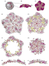
Localization of conserved, non-conserved, and identical amino acids in ARV, MRV, and AqRV core shell protein. The MRV core crystal structure (Reinisch et al., 2000) asymmetric unit (PDB #1EJ6) was assembled into a pentameric vertex aggregate with Viper® (Reddy et al., 2001) and manipulated with Chimera® (Pettersen et al., 2004). (A) Low-resolution, cutaway model of the reovirus core structure (modified from (Dryden et al., 2008) with permission). (B–C) Blow-up of indicated λ1 molecules in “A”; with “C” rotated 90° towards the viewer to better illustrate the decameric organization of 10 λ1 molecules. The λ1 molecules that approach the 5′-axis (designated λ1.5) are shown in salmon and the λ1 molecules that approach the 3′-axis (designated λ1.3) are shown in magenta. (D) Same top view of decameric structure as “C”, but in “D”–“G”, amino acids that are identical in all 6 ARV, MRV, and AqRV sequences (see Fig. 1) are shown in darker versions of each motif color (firebrick and deep pink, respectively), amino acids that represent conservative substitutions (as determined by blossum50 matrix) are shown in lighter versions of each motif color (salmon and hotpink, respectively), and non-conserved amino acids are shown in white and grey, respectively, for λ1.5 and λ1.3. (E) Bottom view of decamer with coloration same as in “D”; in addition, the N-terminal 240 residues, which are visualized only in λ1.3 molecules, are depicted in pale yellow (for non-conservative substitutions), cyan for conservative substitutions, and blue for identical amino acids, respectively. (F) Slabbed section of “D”, approximately as indicated by labelled horizontal line in “B”; note that apparent “hole” in structure is caused by linear transverse section of convex shell. (G) Slabbed section of “E”, approximately as indicated by labelled horizontal line in “B”. (H) Slabbed cross-section through middle of “D”, as indicated by labelled line “H”. (I) Slabbed cross-section through edge of “D”, as indicated by labelled line “I”. The highly conserved C2H2 zinc finger motif is indicated in black (and with large arrow heads) in one λ1.3 molecule, highly conserved residues in putative helicase domains are indicated in green, and highly conserved residues in putative 5′ RNA triphosphatase domains are indicated in gold (and with narrow arrows) in only some molecules. Some highly conserved patches (see text) are indicated in some λ1.5 (circles and hexagons) and λ1.3 molecules (squares and hexagons) in “D”, “H”, and “I”.
Fig. 7
Fig. 7
Localization of conserved, non-conserved, and identical amino acids in ARV, MRV, and AqRV turret proteins. The MRV core crystal structure (Reinisch et al., 2000) asymmetric unit (PDB #1EJ6) was assembled into a pentameric vertex aggregate with Viper® (Reddy et al., 2001), and manipulated with Chimera® (Pettersen et al., 2004). (A) Low-resolution, cutaway model of the reovirus core structure (modified from (Dryden et al., 2008) with permission). (B) Blow-up of indicated λ2 turret in ‘A’, with front-most λ2 molecule in space-filling mode and color-coded according to domain; pink for the N-terminal GTase domain (amino acids 1–380), grey for the extended region (aa. 381–432), yellow for the MTase-1 domain (residues 433–691), green for the spacer domain (aa. 692–805), blue for the MTase-2 domain (aa. 806–1022), and purple for the flap domain (residues 1023–1289); the four other λ2 molecules in the turret are depicted in cyan in wire backbone mode. In “C”–“L”, amino acids that are identical in all six ARV, MRV, and AqRV sequences (see Fig. 4) are shown in darker versions of each domain color (deep pink, dim grey, goldenrod, dark green, blue, and purple, respectively), amino acids that represent conservative substitutions (as determined by blossum50 matrix) are shown in lighter versions of each domain color (hotpink, dark grey, yellow, green, cyan, and plum, respectively), and non-conserved amino acids are shown in white. Highly conserved lysine resides (Lys171 and Lys190 in the MRV sequence, shown to be important for guanylyltransferase activity (Luongo et al., 2000; Luongo, 2002)) are indicated in black, highly conserved MTase-1 residues His521, Asp561, Asp577, and Asp579 (see text) are depicted in magenta, and highly conserved MTase-2 residues Asp827, Gly829, Asp850, and Tyr872 (see text) are shown in orange-red. (C) (top view) and (D) (bottom view) of flaps domain; (E) (top view) and (F) (bottom view) of MTase domains; (H) (top view) and (I) (bottom view) of spacer and extended region domains; and (J) (top view) and (K) (bottom view) of GTase domain. (G and L) Slabbed sections (cut approximately mid-way and viewed from top) of MTase and GTase domains, respectively, to allow better visualization of catalytic sites and indicated highly conserved residues.

Similar articles

Cited by

References

    1. Bartlett JA, Joklik WK. The sequence of the reovirus serotype 3 L3 genome segment which encodes the major core protein lambda 1. Virology. 1988;167:31–37. - PubMed
    1. Benavente J, Martinez-Costas J. Avian reovirus: structure and biology. Virus Res. 2007;123:105–119. - PubMed
    1. Bisaillon M, Bergeron J, Lemay G. Characterization of the nucleoside triphosphate phosphohydrolase and helicase activities of the reovirus lambda1 protein. J Biol Chem. 1997;272:18298–18303. - PubMed
    1. Bisaillon M, Lemay G. Computational sequence analysis of mammalian reovirus proteins. Virus Genes. 1999;18:13–37. - PMC - PubMed
    1. Bodelon G, Labrada L, Martinez-Costas J, Benavente J. The avian reovirus genome segment S1 is a functionally tricistronic gene that expresses one structural and two nonstructural proteins in infected cells. Virology. 2001;290:181–191. - PubMed

Publication types

Associated data

Grants and funding

LinkOut - more resources