

Involvement of human decidual cell-expressed tissue factor in uterine hemostasis and abruption
- PMID: 19720393
- PMCID: PMC2787516
- DOI: 10.1016/j.thromres.2009.07.017
Involvement of human decidual cell-expressed tissue factor in uterine hemostasis and abruption
Abstract
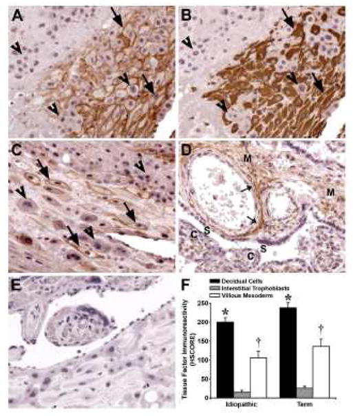
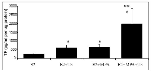
Vascular injury increases access and binding of plasma-derived factor VII to perivascular cell membrane-bound tissue factor (TF). The resulting TF/VIIa complex promotes hemostasis by cleaving pro-thrombin to thrombin leading to the fibrin clot. In human pregnancy, decidual cell-expressed TF prevents decidual hemorrhage (abruption). During placentation, trophoblasts remodel decidual spiral arteries into high conductance vessels. Shallow trophoblast invasion impedes decidual vascular conversion, producing an inadequate uteroplacental blood flow that elicits abruption-related placental ischemia. Thrombin induces several biological effects via cell surface protease activated receptors. In first trimester human DCs thrombin increases synthesis of sFlt-1, which elicits placental ischemia by impeding angiogenesis-related decidual vascular remodeling. During pregnacy, the fibrillar collagen-rich amnion and choriodecidua extracellular matrix (ECM) provides greater than additive tensile strength and structural integrity. Thrombin acts as an autocrine/paracrine mediator that degrades these ECMs by augmenting decidual cell expression of: 1) matrix metalloproteinases and 2) interleukin-8, a key mediator of abruption-associated decidual infiltration of neutrophils, which express several ECM degrading proteases. Among the cell types at the maternal fetal interface at term, TF expression is highest in decidual cells indicating that this TF meets the hemostatic demands of labor and delivery. TF expression in cultured term decidual cells is enhanced by progestin and thrombin suggesting that the maintenance of elevated circulating progesterone provides hemostatic protection and that abruption-generated thrombin acts in an autocrine/paracrine fashion on decidual cells to promote hemostasis via enhanced TF expression.
Figures
Similar articles
-
The role of decidual cells in uterine hemostasis, menstruation, inflammation, adverse pregnancy outcomes and abnormal uterine bleeding.Hum Reprod Update. 2016 Jun;22(4):497-515. doi: 10.1093/humupd/dmw004. Epub 2016 Feb 23. Hum Reprod Update. 2016. PMID: 26912000 Free PMC article. Review.
-
Decidual cell-expressed tissue factor in human pregnancy and its involvement in hemostasis and preeclampsia-related angiogenesis.Ann N Y Acad Sci. 2008 Apr;1127:67-72. doi: 10.1196/annals.1434.013. Ann N Y Acad Sci. 2008. PMID: 18443332 Review.
-
The role of decidualization in regulating endometrial hemostasis during the menstrual cycle, gestation, and in pathological states.Semin Thromb Hemost. 2007 Feb;33(1):111-7. doi: 10.1055/s-2006-958469. Semin Thromb Hemost. 2007. PMID: 17253197 Review.
-
Decidual hemostasis, inflammation, and angiogenesis in pre-eclampsia.Semin Thromb Hemost. 2011 Mar;37(2):158-64. doi: 10.1055/s-0030-1270344. Epub 2011 Mar 2. Semin Thromb Hemost. 2011. PMID: 21370218 Free PMC article.
-
Progestin and thrombin regulate tissue factor expression in human term decidual cells.J Clin Endocrinol Metab. 2009 Jun;94(6):2164-70. doi: 10.1210/jc.2009-0065. Epub 2009 Mar 10. J Clin Endocrinol Metab. 2009. PMID: 19276228 Free PMC article.
Cited by
-
Exploration of the Relationship between Polycystic Ovary Syndrome and Recurrent Pregnancy Loss Based on Bioinformatics.Endocr Metab Immune Disord Drug Targets. 2025;25(7):569-581. doi: 10.2174/0118715303308816240918062247. Endocr Metab Immune Disord Drug Targets. 2025. PMID: 39364875
-
Platelet activation and placenta-mediated adverse pregnancy outcomes: an ancillary study to the Effects of Aspirin in Gestation and Reproduction trial.Am J Obstet Gynecol. 2020 Nov;223(5):741.e1-741.e12. doi: 10.1016/j.ajog.2020.05.026. Epub 2020 May 17. Am J Obstet Gynecol. 2020. PMID: 32434001 Free PMC article.
-
HOXD8/DIAPH2-AS1 epigenetically regulates PAX3 and impairs HTR-8/SVneo cell function under hypoxia.Biosci Rep. 2019 Jan 25;39(1):BSR20182022. doi: 10.1042/BSR20182022. Print 2019 Jan 31. Biosci Rep. 2019. PMID: 30626726 Free PMC article.
-
Impact of blood hypercoagulability on in vitro fertilization outcomes: a prospective longitudinal observational study.Thromb J. 2017 Mar 28;15:9. doi: 10.1186/s12959-017-0131-7. eCollection 2017. Thromb J. 2017. PMID: 28360822 Free PMC article.
-
Inside the Endometrial Cell Signaling Subway: Mind the Gap(s).Int J Mol Sci. 2018 Aug 21;19(9):2477. doi: 10.3390/ijms19092477. Int J Mol Sci. 2018. PMID: 30134622 Free PMC article. Review.
References
-
- Mackman N. Role of tissue factor in hemostasis, thrombosis, and vascular development. Arterioscler Thromb Vasc Biol. 2004;24:1015–1022. - PubMed
-
- Mackman N, Tilley RE, Key NS. Role of the extrinsic pathway of blood coagulation in hemostasis and thrombosis. Arterioscler Thromb Vasc Biol. 2007;27:1687–1693. - PubMed
-
- Tilley R, Mackman N. Tissue factor in hemostasis and thrombosis. Semin Thromb Hemost. 2006;32:5–10. - PubMed
Publication types
MeSH terms
Substances
Grants and funding
LinkOut - more resources
Full Text Sources
Medical
Miscellaneous
