Review
. 2009 Oct 12;364(1531):2809-17.
doi: 10.1098/rstb.2009.0104.

The 'division of labour' model of eye evolution

Affiliations
Review

The 'division of labour' model of eye evolution

Detlev Arendt et al. Philos Trans R Soc Lond B Biol Sci. .

Abstract

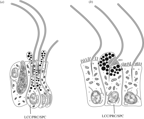
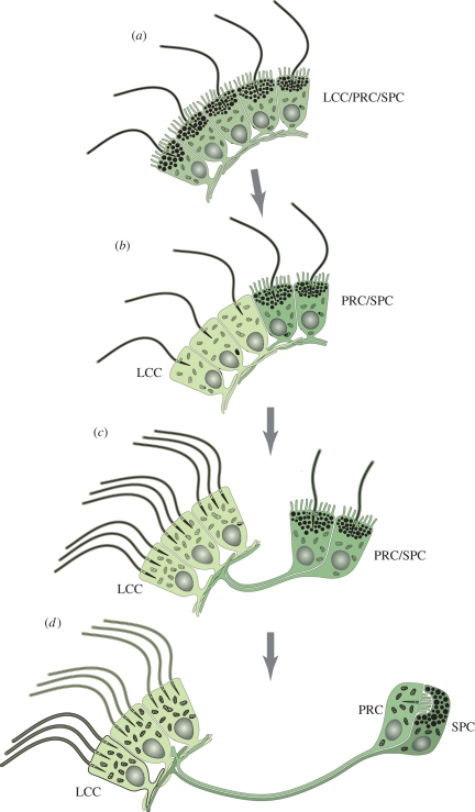
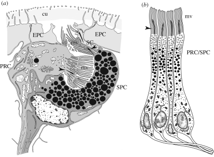
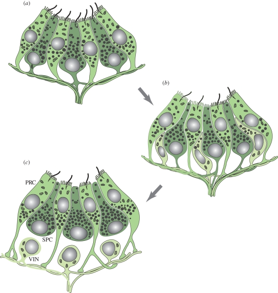
The 'division of labour' model of eye evolution is elaborated here. We propose that the evolution of complex, multicellular animal eyes started from a single, multi-functional cell type that existed in metazoan ancestors. This ancient cell type had at least three functions: light detection via a photoreceptive organelle, light shading by means of pigment granules and steering through locomotor cilia. Located around the circumference of swimming ciliated zooplankton larvae, these ancient cells were able to mediate phototaxis in the absence of a nervous system. This precursor then diversified, by cell-type functional segregation, into sister cell types that specialized in different subfunctions, evolving into separate photoreceptor cells, shading pigment cells (SPCs) or ciliated locomotor cells. Photoreceptor sensory cells and ciliated locomotor cells remained interconnected by newly evolving axons, giving rise to an early axonal circuit. In some evolutionary lines, residual functions prevailed in the specialized cell types that mirror the ancient multi-functionality, for instance, SPCs expressing an opsin as well as possessing rhabdomer-like microvilli, vestigial cilia and an axon. Functional segregation of cell types in eye evolution also explains the emergence of more elaborate photosensory-motor axonal circuits, with interneurons relaying the visual information.


Figures

Figure 1.
Figure 1.
Simple multi-functional PRCs (LCC/PRC/SPC) with motile cilia, sensory microvilli and shading pigment granules. (a) In the demosponge larva (Amphimedon), shading pigment prevents admittance of light to light-sensitive part of the cells from several directions. The cilia is active in phototactic steering. Note the absence of microvilli. After Leys & Degnan (2001) and Maldonado et al. (2003). (b) Rhabdomeric PRC in the planula larva of the cubozoan cnidarian Tripedalia; rhabdomeric microvilli (mv) shaded from various directions (Nordstrom et al. 2003).
Figure 2.
Figure 2.
The axonal scaffold of the larval brain of P. dumerilii at 48 hpf. Rhabdomeric PRCs (red) projecting to the ciliary girdle, where they locally alter the strength of ciliary beating (Jékely et al. 2008). Grey colour indicates axons and cilia labelled by an antibody directed against acetylated tubulin. Blue dots have been added following phalloidin stainings that specifically label the actin filaments of microvilli making up the rhabdomeres. Photograph courtesy of G. Jékely.
Figure 3.
Figure 3.
Evolution of two-celled rhabdomeric larval eyes mediating phototaxis, according to the division of labour model. Multi-functional locomotor-photosensory precursor cells evolve into LCCs by cell-type functional segregation. (a) Precursor LCC/PRC/SPC combining a locomotor cilium, photosensory rhabdomeric microvilli and shading pigment granules. (b) A subset of cells loses photosensitivity and shading pigment to specialize on locomotion (LCC, light green). (c) The LCCs become multiciliated. A spatially separate ‘eye’ comprising two combined PRC/SPC remains connected to the multiciliated cells via gradually extending cellular processes that evolve into axons. (d) Functional segregation of PRCs (green) and SPCs (dark green). For further explanations, see text.
Figure 4.
Figure 4.
(a) Two-celled eye of Saccocirrus papillocercus after transmission electron microscopy observations. PRC and SPC are embedded in the layer of epidermal cells (EPCs); both cells with microvilli (mv) in rhabdomeric arrangement, vestigial cilia (arrowheads) and axonal processes; a few scattered electron-dense vesicles in PRC may comprise shading pigment. Note additional sensory cell (SC) adjacent to pigment cell. cu, cuticle. (b) Adult eye retina cells of Patella exclusively composed of combined PRC/SPC. Note that each cell possesses a cilium (arrowhead) and sends out an axon. mv, microvilli (adapted from Marshall & Hodgson 1990).
Figure 5.
Figure 5.
Evolution of visual interneurons according to the division of labour model. (a) Pigment cup eye composed of partially segregated photoreceptor (green) and SPCs (dark green). Note that both cell types bear sensory microvilli and shading pigment granules, but in different quantity. Together, the pigment granules of both cell types form the eye cup. Both cell types also send out axons. (b) A subset of cells loses photosensitivity and shading pigment to specialize on information integration (light green). The pigment cells lose the cilia. (c) Evolution of fully developed unipolar visual interneurons (VIN) that retain axonal connections to multiple PRC. The SPCs (dark green) have specialized on their shading function and lost the sensory microvilli as well as basal axons. Note that PRCs axons connect to VIN exclusively. For further explanations, see text.

References

    1. Arendt D.2003Evolution of eyes and photoreceptor cell types. Int. J. Dev. Biol. 47, 563–571 - PubMed
    1. Arendt D.2008The evolution of cell types in animals: emerging principles from molecular studies. Nat. Rev. Genet. 9, 868–882 (doi:10.1038/nrg2416) - DOI - PubMed
    1. Arendt D., Wittbrodt J.2001Reconstructing the eyes of Urbilateria. Phil. Trans. R. Soc. Lond. B 356, 1545–1563 (doi:10.1098/rstb.2001.0971) - DOI - PMC - PubMed
    1. Arneson A. C., Cutress C. E.1976Life history of Carybdea alata Reynaud, 1830 (Cubomedusae). In Coelenterate ecology and behavior (ed. Mackie G. O.), New York, NY: Plenum
    1. Bartolomaeus T.1992aUltrastructure of the photoreceptors in certain larvae of the Annelida. Microfauna Mar. 7, 191–214

MeSH terms