Review
. 2009 Oct 12;364(1531):2849-65.
doi: 10.1098/rstb.2009.0050.

The evolution of irradiance detection: melanopsin and the non-visual opsins

Affiliations
Review

The evolution of irradiance detection: melanopsin and the non-visual opsins

Stuart N Peirson et al. Philos Trans R Soc Lond B Biol Sci. .

Abstract

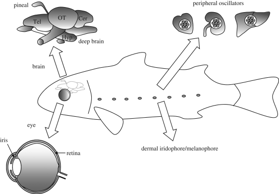
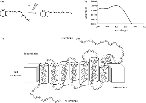
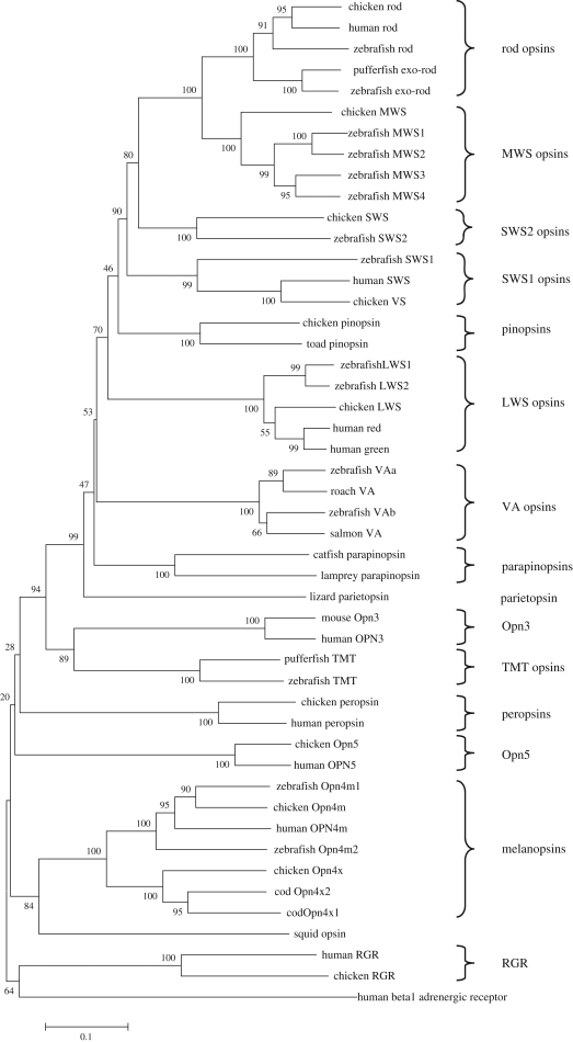
Circadian rhythms are endogenous 24 h cycles that persist in the absence of external time cues. These rhythms provide an internal representation of day length and optimize physiology and behaviour to the varying demands of the solar cycle. These clocks require daily adjustment to local time and the primary time cue (zeitgeber) used by most vertebrates is the daily change in the amount of environmental light (irradiance) at dawn and dusk, a process termed photoentrainment. Attempts to understand the photoreceptor mechanisms mediating non-image-forming responses to light, such as photoentrainment, have resulted in the discovery of a remarkable array of different photoreceptors and photopigment families, all of which appear to use a basic opsin/vitamin A-based photopigment biochemistry. In non-mammalian vertebrates, specialized photoreceptors are located within the pineal complex, deep brain and dermal melanophores. There is also strong evidence in fish and amphibians for the direct photic regulation of circadian clocks in multiple tissues. By contrast, mammals possess only ocular photoreceptors. However, in addition to the image-forming rods and cones of the retina, there exists a third photoreceptor system based on a subset of melanopsin-expressing photosensitive retinal ganglion cells (pRGCs). In this review, we discuss the range of vertebrate photoreceptors and their opsin photopigments, describe the melanopsin/pRGC system in some detail and then finally consider the molecular evolution and sensory ecology of these non-image-forming photoreceptor systems.


Figures

Figure 1.
Figure 1.
Photoreceptive sites in the vertebrates. As well as the classical photoreceptors within the retina of the lateral eye, direct photoreception in the isolated iris has also been described. In non-mammalian species the pineal complex also contains photoreceptors, and deep brain photoreceptors may also occur. Dermal photoreception has been described in amphibians and fish. Finally, in the zebrafish peripheral tissues have been shown to be able to entrain their molecular oscillators directly to light. See text for further details.
Figure 2.
Figure 2.
Structure and function of vertebrate photopigments. Vertebrate photopigments consist of an isomer of vitamin A, retinaldehyde, bound to an opsin protein. (a) The primary step in phototransduction is the absorption of a photon of light (hv) by the 11-cis isomer of retinal resulting in isomerization to the all-trans form. (b) All vitamin A/opsin-based photopigments have a characteristic absorption spectrum which can be used as a ‘spectral fingerprint’ to determine the photopigment mediating a given biological response. (c) Opsins consist of a single polypeptide chain forming seven α-helical transmembrane regions connected by cytoplasmic and extracellular loops. The intracellular domains mediate G-protein interactions. The retinal binding site (K) is indicated in the 7th transmembrane domain. Structure based on that of Palczewski et al. 2000.
Figure 3.
Figure 3.
Phylogenetic tree showing the relationship of the various classes of vertebrate opsins. The entire amino acid sequences were aligned using ClustalW (Higgins et al. 1996) and the tree was generated by the neighbour joining method (Saitou & Nei 1987) using the MEGA4 program (Tamura et al. 2007). Branch confidence levels (% based on 1000 bootstrap replicates) are marked. Scale bar indicates substitutions per site. The human beta 1 adrenergic receptor was used as an outgroup. The major classes of the vertebrate opsins are indicated by parentheses on the right-hand side. The analysis reveals that the exo-rod opsins are a duplication of the rod opsins and that pinopsin has arisen by a duplication of the cone opsins. To date there is only one sequence for parietopsin (see text for details). Opn3 and TMT, both expressed in multiple tissues, seem to share a common ancestor. The Opn4 sequences now quite clearly consist of two families, the mammalian-like ‘m’ form and the Xenopus-like ‘x’ form. Interestingly the opsin from the invertebrate squid also included in the analysis clades with the melanopsin sequences, adding credence to the argument that melanopsin is ‘invertebrate’-like. Finally RGR is the least similar to the visual opsins. Accession numbers: Chicken: rod D00702; LWS M62903; MWS M92038; SWS M92037; versus M92039; RGR AY339627; peropsin AY339626; pinopsin U15762; Opn4m AY036061; Opn4x AY882944; Opn5 XM_001130743. Human: rod NM_000539; red NM_020061; green NM_000513; SWS NM_001708; RGR NM_002921; peropsin NM_006583; OPN3 NM_014322; OPN4M NM_033282; OPN5 NM_181744; beta 1 adrenergic receptor NM_000684. Zebrafish: rod NM_131084; LWS1, NM_001002443; LWS2, NM_131175; MWS1, NM_131253; MWS2, NM_182891; MWS3, NM_182892; MWS4, NM_131254; SWS1, NM_131319; SWS2, NM_131192; exo-rod, AB025312; VA1, AB035276; VA2, AY996588; TMT, AF349947; Opn4m1, AY882945; Opn4m2, AY078161. Pufferfish: exo-rod, AF201472; TMT, AF402774. Salmon VA AF001499. Roach VA AY116411. Catfish parapinopsin AF028014. Lamprey parapinopsin AB116380. Lizard parietopsin DQ100320. Toad pinopsin AF200433. Mouse Opn3 NM_010098. Cod Opn4x1 AF385823. Opn4x2 AY126448. Squid opsin, P09241.

References

    1. Altimus C. M., Guler A. D., Villa K. L., McNeill D. S., Legates T. A., Hattar S.2008Rods–cones and melanopsin detect light and dark to modulate sleep independent of image formation. Proc. Natl Acad. Sci. USA 105, 19 998–20 003 (doi:10.1073/pnas.0808312105) - DOI - PMC - PubMed
    1. Arendt J.1998Melatonin and the pineal gland: influence on mammalian seasonal and circadian physiology. Rev. Reprod. 3, 13–22 (doi:10.1530/ror.0.0030013) - DOI - PubMed
    1. Arendt D.2003Evolution of eyes and photoreceptor cell types. Int. J. Dev. Biol. 47, 563–571 - PubMed
    1. Aschoff J.1984Circadian timing. Ann. N. Y. Acad. Sci. 423, 442–468 (doi:10.1111/j.1749-6632.1984.tb23452.x) - DOI - PubMed
    1. Barlow H. B.1957Purkinje shift and retinal noise. Nature 179, 255–256 (doi:10.1038/179255b0) - DOI - PubMed

LinkOut - more resources