Review
. 2009 Oct 12;364(1531):2867-80.
doi: 10.1098/rstb.2009.0077.

Evolution of vertebrate rod and cone phototransduction genes

Affiliations
Review

Evolution of vertebrate rod and cone phototransduction genes

Dan Larhammar et al. Philos Trans R Soc Lond B Biol Sci. .

Abstract

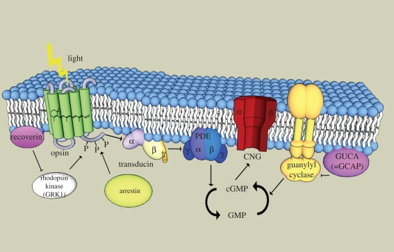
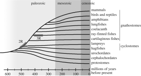
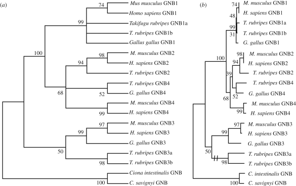
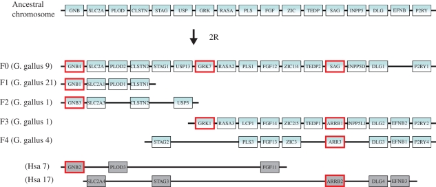
Vertebrate cones and rods in several cases use separate but related components for their signal transduction (opsins, G-proteins, ion channels, etc.). Some of these proteins are also used differentially in other cell types in the retina. Because cones, rods and other retinal cell types originated in early vertebrate evolution, it is of interest to see if their specific genes arose in the extensive gene duplications that took place in the ancestor of the jawed vertebrates (gnathostomes) by two tetraploidizations (genome doublings). The ancestor of teleost fishes subsequently underwent a third tetraploidization. Our previously reported analyses showed that several gene families in the vertebrate visual phototransduction cascade received new members in the basal tetraploidizations. We here expand these data with studies of additional gene families and vertebrate species. We conclude that no less than 10 of the 13 studied phototransduction gene families received additional members in the two basal vertebrate tetraploidizations. Also the remaining three families seem to have undergone duplications during the same time period but it is unclear if this happened as a result of the tetraploidizations. The implications of the many early vertebrate gene duplications for functional specialization of specific retinal cell types, particularly cones and rods, are discussed.


Figures

Figure 1.
Figure 1.
Schematic outline of the phototransduction cascade in a vertebrate cone. All of the components are shown within or near the same membrane, the cell-surface membrane, as is assumed to have been the case in the ancestor of cones and rods (rods subsequently evolved an internalization mechanism for parts of the membrane to form intracellular discs whose membranes harbour some of the phototransduction components including rhodopsin). Upon absorption by a photon, 11-cis-retinal within the opsin is transformed to all-trans-retinal. This activates the opsin, which in turn activates the transducin (a G protein) consisting of three subunits (α, β and γ). Transducin in turn activates phophodiesterase 6 (PDE), which forms a dimer with α and β (or two α subunits), inhibited by two γ subunits. Active PDE hydrolyzes cGMP to GMP. cGMP keeps the cyclic nucleotide ion channel (CNG, a tetramer with α and β subunits) open. New cGMP is generated by guanylyl cyclase (GC), which is regulated by a GC-activating protein (GUCA or GCAP). Rhodopsin kinase (GRK) deactivates the opsin by phosphorylation, and is itself regulated by recoverin. Arrestin binds to the phosphorylated opsin to reduce its signalling to the G protein. GRK is anchored to the membrane and arrestin undergoes translocation to the membrane on activation (not shown in the figure).
Figure 2.
Figure 2.
Evolutionary tree for the different classes of gnathostomes (jawed vertebrates) in relation to cyclostomes (lampreys and hagfishes), urochordates (tunicates) and cephalochordates (lancelets). It is still unclear when the two genome-wide duplications (2R) took place in relation to the split of the cyclostomes from the gnathostomes. It is also still unclear whether lampreys and hagfishes are monophyletic or paraphyletic, which is why their deep branch is dashed in the figure. A third independent genome duplication (3R) took place in the teleost lineage.
Figure 3.
Figure 3.
Chromosomal locations of vertebrate visual opsin gene family members in the human genome (Hsa for Homo sapiens) and postulated positions for the lost ancestral blue and green opsins. The proposed position for the ancient green opsin (RHO2) is confirmed by the present location of the orthologue in the chicken genome in a region with conserved synteny with Hsa1. Because phylogenetic trees for vertebrate visual opsins have OPN1LW and OPN1SW as the most basal divergence (Bowmaker 2008), we postulate that there was an ancestral gene pair before the chromosome quadruplication. However, other scenarios are possible. This hypothetical chromosome duplication scenario needs to be investigated further by studies in genomes of representatives from additional vertebrate classes. The transducin α subunit (GNAT) involved in phototransduction is located in the same paralogon, with GNAT1 expressed in rods, and GNAT2 in cones.
Figure 4.
Figure 4.
Phylogenetic trees for the G-protein β subunit family GNB1–4. The species included are two mammals (human and mouse), one bird (chicken) and a pufferfish (Takifugu rubripes). As outgroup the GNB gene of the tunicates Ciona intestinalis and Ciona savignyi were used. The trees were calculated using the neighbour-joining method as implemented in Mega 4.0 (Tamura et al. 2007) with default settings. Panel (a) shows the obtained tree collapsed for nodes with less than 50 per cent bootstrap support. Panel (b) shows the same tree with branch lengths representing evolutionary distance. The two T. rubripes genes GNB3a and GNB3b have evolved at a much higher rate than all of the other sequences and their common branch has been shortened as shown by the double dash in panel (b).
Figure 5.
Figure 5.
Chromosomal locations of vertebrate phototransduction gene families GNB1/2/3/4, GRK1/7 and Arrestins/SAG together with several adjacent gene families in the chicken genome (Gga for Gallus gallus) and in the human genome (Hsa for Homo sapiens) in paralogon F as named by (Nakatani et al. 2007). The phototransduction genes are highlighted by red boxes. Rods express GNB1, GRK1 and SAG, and cones express GNB3, GRK7 and ARR3. The top chromosome shows the deduced ancestral chromosome that gave rise to the duplicated chromosomal segments in gnathostomes, represented here by the chicken. The five chicken chromosomes together account for three copies of the ancestral chromosome (further analyses in additional species are required to say whether segment F1 shall be combined with F3 or F4). As the GNB2 gene and the ARRB2 gene are missing in the chicken genome database, the human orthologues and their chromosomal regions are shown instead. Numerous rearrangements have occurred after the chromosome quadruplication. Note that the gene order has been re-shuffled to highlight the similarity between the chromosomes.

Similar articles

Cited by

References

    1. Abi-Rached L., Gilles A., Shiina T., Pontarotti P., Inoko H.2002Evidence of en bloc duplication in vertebrate genomes. Nat. Genet. 31, 100–105 (doi:10.1038/ng855) - DOI - PubMed
    1. Ames J. B., Ikura M.2002Structure and membrane-targeting mechanism of retinal Ca2+-binding proteins, recoverin and GCAP-2. Adv. Exp. Med. Biol. 514, 333–348 - PubMed
    1. Applebury M. L., et al. 2000The murine cone photoreceptor: a single cone type expresses both S and M opsins with retinal spatial patterning. Neuron 27, 513–523 (doi:10.1016/S0896-6273(00)00062-3) - DOI - PubMed
    1. Arendt D.2008The evolution of cell types in animals: emerging principles from molecular studies. Nat. Rev. Genet. 9, 868–882 (doi:10.1038/nrg2416) - DOI - PubMed
    1. Balcueva E. A., Wang Q., Hughes H., Kunsch C., Yu Z., Robishaw J. D.2000Human G protein gamma(11) and gamma(14) subtypes define a new functional subclass. Exp. Cell Res. 257, 310–319 (doi:10.1006/excr.2000.4893) - DOI - PubMed

Publication types

LinkOut - more resources