Review
. 2009 Oct 12;364(1531):2881-95.
doi: 10.1098/rstb.2009.0051.

Evolution of opsins and phototransduction

Affiliations
Review

Evolution of opsins and phototransduction

Yoshinori Shichida et al. Philos Trans R Soc Lond B Biol Sci. .

Abstract

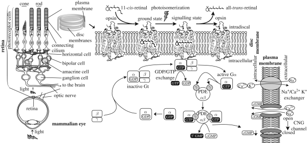
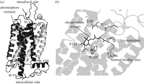
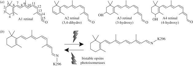
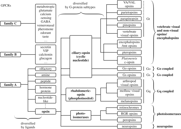
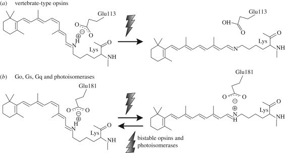
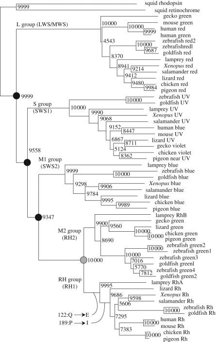
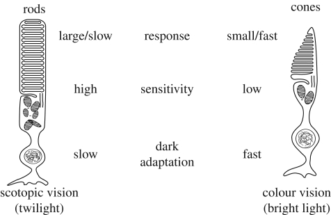
Opsins are the universal photoreceptor molecules of all visual systems in the animal kingdom. They can change their conformation from a resting state to a signalling state upon light absorption, which activates the G protein, thereby resulting in a signalling cascade that produces physiological responses. This process of capturing a photon and transforming it into a physiological response is known as phototransduction. Recent cloning techniques have revealed the rich and diverse nature of these molecules, found in organisms ranging from jellyfish to humans, functioning in visual and non-visual phototransduction systems and photoisomerases. Here we describe the diversity of these proteins and their role in phototransduction. Then we explore the molecular properties of opsins, by analysing site-directed mutants, strategically designed by phylogenetic comparison. This site-directed mutant approach led us to identify many key features in the evolution of the photoreceptor molecules. In particular, we will discuss the evolution of the counterion, the reduction of agonist binding to the receptor, and the molecular properties that characterize rod opsins apart from cone opsins. We will show how the advances in molecular biology and biophysics have given us insights into how evolution works at the molecular level.


Figures

Figure 1.
Figure 1.
A diagram showing the mechanism of phototransduction in mammalian eyes. Light is captured by two specialized morphologically distinct photoreceptor cells derived from neurons: rods and cones that have the same molecular mechanism. Opsins in these cells absorb photons and form a signalling state, which can bind to and activate the G protein by catalysing the exchange of GDP to GTP. The GTP-bound Gα dissociates from Gβγ exposing its active site. Activated Gα binds to its effector, PDE (cyclic nucleotide phosphodiesterase), and activates it. PDE breaks the phosphodiester bond of cGMP producing 5′GMP, and the decrease in the concentration of cGMP causes CNG (cyclic nucleotide gated) channels to close, which creates a hyperpolarization response in the photoreceptor cells. Light-activated rhodopsin is thermally unstable and the chromophore eventually detaches from the opsin. The hyperpolarization of the membrane potential of the photoreceptor cell modulates the release of neurotransmitters to downstream cells. The light signal is transmitted through different cells, finally reaching ganglion cells which form the optic nerve and project to the brain.
Figure 2.
Figure 2.
Molecular structure of rhodopsin. (a) Cartoon representation of the atomic model of rhodopsin, consisting of seven transmembrane helices, an eighth helix at the intracellular side and parallel to the membrane, and the chromophore shown as spheres. (b) A close-up of the structure of the chromophore and the spacial location of some of the amino acids that characterize rhodopsin, discussed in this article. Molecular graphics representations were created using PyMol (DeLano Scientific LLC).
Figure 3.
Figure 3.
(a) Differences in A1, A2, A3 and A4 retinals used by opsins. (b) Photoisomerization of the retinal. In opsins, the retinal is covalently bound to a lysine located at H7, and the isomerization is sterospecific from 11-cis to all-trans. Some opsins are bistable and the photoisomerized all-trans-retinal can be reconverted to 11-cis-retinal by the absorption of a second photon. Also there are photoisomerases, opsins that bind all-trans-retinal and form 11-cis-retinal.
Figure 4.
Figure 4.
Schematic representation of the phylogenic relationship of opsins. Opsins belong to the family-A GPCRs, and they can be roughly subdivided into ciliary opsins, rhadbomeric opsins and photoisomerases. The ciliary opsins are characterized by their expression in ciliary photoreceptor cells and cyclic nucleotide signalling cascade. On the other hand, rhabdomeric opsins are expressed in rhabdomeric photoreceptor cells and have phosphoinositol signalling cascade. Finally, photoisomerases comprises proven and putative stereospecific photoisomerases.
Figure 5.
Figure 5.
The counterion of opsins. (a) Vertebrate visual and non-visual opsins have a negatively charged glutamic acid at 113 in H3 that functions as the counterion for the positive charge of the protonated retinal Schiff base. (b) The counterion of Go/Gs/Gq opsins and photoisomerases is E181.
Figure 6.
Figure 6.
Phylogenetic tree of vertebrate visual opsins constructed by NJ (numbers at the nodes indicate the bootstrap values of 10 000 replicates). Five distinctive groups, which correspond well with their spectral sensitivities, can be identified: the UV or violet light-sensitive S (or SWS1) group, the blue light-sensitive M1 (or SWS2) group, the green light-sensitive M2 (or RH2) group, the red or green light-sensitive L (or LWS/MWS) group and the scotopic vision RH (or RH1) group. Note that the tree does not necessarily reflect the phylogenic relationship of organisms. Black circle, colour vision; grey circle, twilight vision.
Figure 7.
Figure 7.
Response profiles of rods and cones. Rods are characterized by a large and slow response, high sensitivity and a slow dark adaptation. On the other hand, cones are characterized by a small and fast response, low sensitivity and a fast dark adaptation. These response profiles originate from the functional proteins in them.

Similar articles

Cited by

References

    1. Arendt D.2003Evolution of eyes and photoreceptor cell types. Int. J. Dev. Biol. 47, 563–571 - PubMed
    1. Arendt D., Tessmar-Raible K., Snyman H., Dorresteijn A. W., Wittbrodt J.2004Ciliary photoreceptors with a vertebrate-type opsin in an invertebrate brain. Science 306, 869–871 (doi:10.1126/science.1099955) - DOI - PubMed
    1. Barnard A. R., Hattar S., Hankins M. W., Lucas R. J.2006Melanopsin regulates visual processing in the mouse retina. Curr. Biol. 16, 389–395 (doi:10.1016/j.cub.2005.12.045) - DOI - PubMed
    1. Beck M., Sakmar T. P., Siebert F.1998Spectroscopic evidence for interaction between transmembrane helices 3 and 5 in rhodopsin. Biochemistry 37, 7630–7639 (doi:10.1021/bi9801560) - DOI - PubMed
    1. Blackshaw S., Snyder S. H.1997Parapinopsin, a novel catfish opsin localised to the parapineal organ, defines a new gene family. J. Neurosci. 17, 8083–8092 - PMC - PubMed

Publication types

LinkOut - more resources