Review
. 2009 Oct 12;364(1531):2925-40.
doi: 10.1098/rstb.2009.0099.

The evolution of early vertebrate photoreceptors

Affiliations
Review

The evolution of early vertebrate photoreceptors

Shaun P Collin et al. Philos Trans R Soc Lond B Biol Sci. .

Abstract

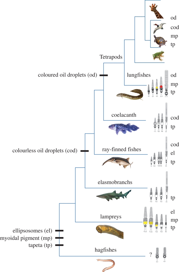
Meeting the challenge of sampling an ancient aquatic landscape by the early vertebrates was crucial to their survival and would establish a retinal bauplan to be used by all subsequent vertebrate descendents. Image-forming eyes were under tremendous selection pressure and the ability to identify suitable prey and detect potential predators was thought to be one of the major drivers of speciation in the Early Cambrian. Based on the fossil record, we know that hagfishes, lampreys, holocephalans, elasmobranchs and lungfishes occupy critical stages in vertebrate evolution, having remained relatively unchanged over hundreds of millions of years. Now using extant representatives of these 'living fossils', we are able to piece together the evolution of vertebrate photoreception. While photoreception in hagfishes appears to be based on light detection and controlling circadian rhythms, rather than image formation, the photoreceptors of lampreys fall into five distinct classes and represent a critical stage in the dichotomy of rods and cones. At least four types of retinal cones sample the visual environment in lampreys mediating photopic (and potentially colour) vision, a sampling strategy retained by lungfishes, some modern teleosts, reptiles and birds. Trichromacy is retained in cartilaginous fishes (at least in batoids and holocephalans), where it is predicted that true scotopic (dim light) vision evolved in the common ancestor of all living gnathostomes. The capacity to discriminate colour and balance the tradeoff between resolution and sensitivity in the early vertebrates was an important driver of eye evolution, where many of the ocular features evolved were retained as vertebrates progressed on to land.


Figures

Figure 1.
Figure 1.
Schematic representations of the spectral sensitivities of the visual pigments of the different photoreceptor types found in early vertebrates. (a) The downstream and (b) upstream migrants of the Southern Hemisphere lamprey, G. australis. The long-wavelength shift (from 492 to 552 nm) of the MWS pigment is caused by the replacement of the yellow myoidal pigment with an ellipsosome. (c) The river lamprey, L. fluviatilis. (d) The sea lamprey, P. marinus. (e) The Southern Hemisphere lamprey, M. mordax. (f) The giant shovelnose ray, G. typus. (g) The juvenile stage of the Australian lungfish, N. forsteri. (h) The adult stage of N. forsteri. Note the loss of the UVS pigment in the adult lungfish. Data predominantly based on microspectrophotometric (MSP) analyses but spectrophotometric analysis of recombinant pigments regenerated with 11-cis-retinal have also been included where MSP was not possible. λmax values (indicated) were obtained from Govardovskii & Lychakov (1984); Hárosi & Kleinschmidt (1993); Collin et al. (2003b, 2004) and Hart et al. (2004, 2008). The dashed lines in (g) and (h) indicate the long-wavelength shift in absorbance after taking into account the intracellular filtering produced by the oil droplets and myoidal pigment.
Figure 2.
Figure 2.
Phylogeny of visual pigment (opsin) gene families (LWS, SWS1, SWS2, RhB/Rh2 and RhA/Rh1) in vertebrates. The tree was generated by using a Bayesian probabilistic inference method with a Metropolis Markov chain Monte Carlo algorithm. A general time-reversal model was used with posterior probability values (represented as a percentage) indicated at the base of each node. The scale bar indicates the number of nucleotide substitutions per site. The fruitfly Rh4 was used as an outgroup.
Figure 3.
Figure 3.
Axial view of the retinal photoreceptor array in a range of early vertebrates viewed using Nomarski optics. (a) Upstream migrant of the Southern Hemisphere lamprey, G. australis (Agnatha). Photoreceptors are differentiated based on the presence of an ellipsosome (large circular profiles), the presence or absence of an orange/yellow, myoidal short wavelength-absorbing pigment and size. (b) Downstream migrant of the Southern Hemisphere lamprey, M. mordax (Agnatha). This species has only one photoreceptor type containing an ellipsosome arranged within a tightly packed hexagonal array and surrounded by a tapetum housed within the retinal pigment epithelial cells. (c) Epaulette shark, Hemiscyllium ocellatum (Chondrichthyes, Elasmobranchii). This rod-rich retina has a low proportion of cones differentiated on the basis of their small size and refractive properties (Litherland & Collin 2008). (d) Australian lungfish, N. forsteri (Osteichthyes, Dipnoi). Large red oil droplets and orange myoidal pigment (arrowheads) are interspersed with large rods and smaller cones containing clear oil droplets. Scale bars: (a) 20 µm; (b) 25 µm; (c) 5 µm; (d) 35 µm.
Figure 4.
Figure 4.
Schematic representation of the complement of photoreceptor types in early vertebrates based on morphological criteria. (a) The single receptor type in the lamprey, M. mordax (Mordaciidae). (b) The long and short receptors in the Northern Hemisphere (holarctic) lampreys (Petromyzontidae). (c) The five receptor types in the lamprey, G. australis (downstream migrant) (Geotriidae). Note the inclusion of yellow myoidal pigment in three of the receptor types. (d) The four receptor types in the giant shovelnose ray, G. typus (Rhinobatidae). (e) The five receptor types in juvenile Australian lungfish, N. forsteri (Ceratodontidae). Note the yellow myoidal pigment and both red and colourless oil droplets. Receptor identities have been based on a range of morphological, spectral and molecular studies; C/R, cone/rod hybrid or features that make it difficult to differentiate; C, cone; R, rod; dm, distended mitochondria; e, ellipsosome; m, mitochondria within the inner segment; n, nucleus; od, oil droplet; os, outer segment; p, paraboloid; yp (1 and 2), orange/yellow pigment.
Figure 5.
Figure 5.
Phylogenetic schematic of the major vertebrate groups showing the evolution of tapeta (tp), myoidal short wavelength-absorbing pigment (mp), ellisosomes (el), colourless oil droplets (cod) and coloured oil droplets (od). Black bars represent the first appearance of each of the morphological traits. The schematic drawings of the photoreceptors on the right-hand side depict the complement of receptor types within at least one species within that vertebrate class/group. The lists of visual traits adjacent to these drawings indicate that at least one species possesses this feature. If a particular feature is not listed for a particular clade, it has not been identified (to our knowledge) in any species within that class/group. The phylogeny is based on that of Meyer & Zardoya (2003). At present, the relationships between hagfishes and lampreys and between lungfishes and the coelacanth are considered polytomies but have been depicted here as being monophyletic based on other visual characters.

Similar articles

Cited by

References

    1. Arrese C. A., Hart N. S., Thomas N., Beazley L. D., Shand J.2002Trichromacy in Australian marsupials. Curr. Biol. 12, 657–660 (doi:10.1016/S0960-9822(02)00772-8) - DOI - PubMed
    1. Avery J. A., Bowmaker J. K.1982Visual pigments in the four-eyed fish. Anableps anableps. Nature 298, 62–64 (doi:10.1038/298062a0) - DOI
    1. Bailes H. J., Robinson S. R., Trezise A. E. O., Collin S. P.2006Morphology, characterisation and distribution of retinal photoreceptors in the Australian lungfish Neoceratodus forsteri (Krefft, 1870). J. Comp. Neurol. 494, 381–397 (doi:10.1002/cne.20809) - DOI - PubMed
    1. Bailes H. J., Davies W. L., Trezise A. E. O., Collin S. P.2007Visual pigments in a living fossil, the Australian lungfish Neoceratodus forsteri. BMC Evol. Biol. 7, 200 (doi:10.1186/1471–2148–7-200) - DOI - PMC - PubMed
    1. Barbour H. R., Archer M. A., Hart N. S., Thomas N., Dunlop S. A., Beazley L. D., Shand J.2002Retinal characteristics of the ornate dragon lizard, Ctenophorous ornatus. J. Comp. Neurol. 450, 334–344 (doi:10.1002/cne.10308) - DOI - PubMed

Publication types

LinkOut - more resources