. 2009 Nov;77(11):4771-82.
doi: 10.1128/IAI.00850-09. Epub 2009 Aug 31.

Yersinia pseudotuberculosis virulence determinants invasin, YopE, and YopT modulate RhoG activity and localization

Affiliations

Yersinia pseudotuberculosis virulence determinants invasin, YopE, and YopT modulate RhoG activity and localization

Sina Mohammadi et al. Infect Immun. 2009 Nov.

Abstract

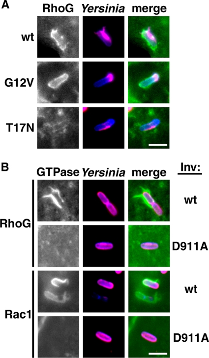
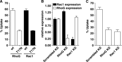
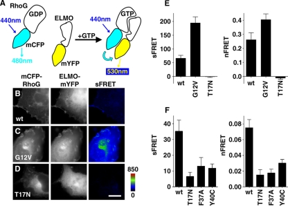
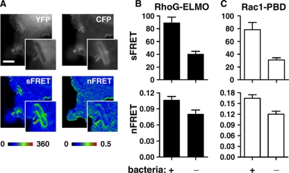
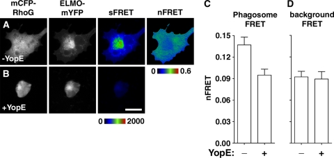
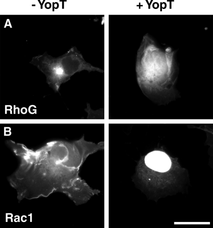
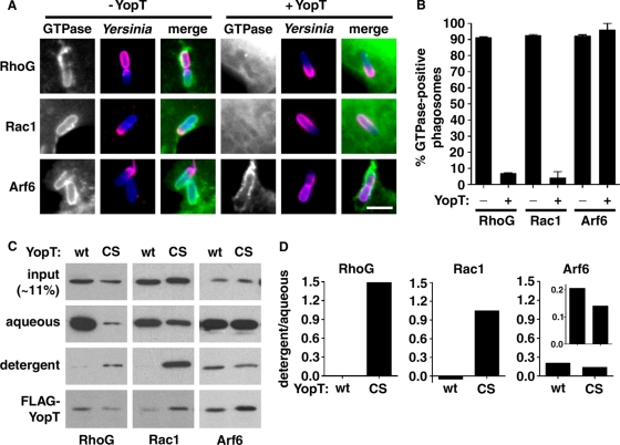
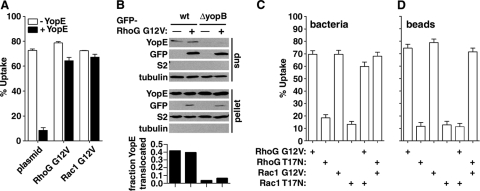
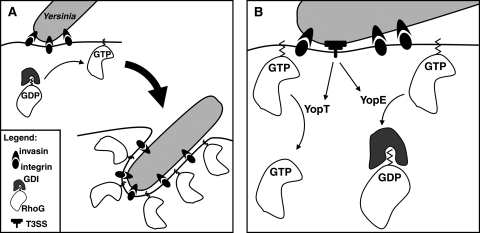
The Yersinia pseudotuberculosis surface protein invasin binds to multiple beta1 integrins with high affinity, leading to misregulation of Rac1 activity. Upon host cell binding, alteration of Rho GTPase activity results from the action of several Yersinia outer proteins (Yops) that are translocated into the cytoplasm. We report here that three virulence determinants encoded by Y. pseudotuberculosis manipulate the Rho GTPase RhoG. Y. pseudotuberculosis binding to cells caused robust recruitment of RhoG to the site of attachment, which required high-affinity invasin-beta1 integrin association. Furthermore, inactivation of RhoG significantly reduced the efficiency of invasin-mediated bacterial internalization. To investigate the activation state of RhoG, a fluorescence resonance energy transfer-based activation biosensor was developed and used to show distinct spatial activation of RhoG at the site of bacterial attachment. The biosensor was also used to show efficient RhoG inactivation by Y. pseudotuberculosis YopE, a potent Rho GTPase activating protein. Additionally, RhoG mislocalization by the prenylcysteine endoprotease YopT was demonstrated by two independent assays. Functional bacterial uptake experiments demonstrated that RhoG activation can bypass a deficit in Rac1 activity. Interestingly, increasing the size of the particle gave results more consistent with a linear pathway, in which RhoG acts as an upstream activator of Rac1, indicating that increased surface area introduces constraints on the signaling pathways required for efficient internalization. Taken together, these data demonstrate the misregulation of RhoG by multiple Y. pseudotuberculosis virulence determinants. Since RhoG is imperative for proper neutrophil function, this misregulation may represent a unique mechanism by which Yersinia species dampen the immune response.


Figures

FIG. 1.
FIG. 1.
RhoG localizes to nascent Yersinia-containing phagosomes in an invasin-dependent manner. (A) Wild-type and constitutively active but not dominant-negative RhoG proteins localize to nascent phagosomes. COS1 cells expressing wild-type (wt), constitutively active (G12V), or dominant-negative (T17N) RhoG constructs (GFP fusions) were incubated briefly (∼20 min) with Y. pseudotuberculosis YPIII(P). Localization at nascent phagosomes was visualized by fluorescence microscopy (see Materials and Methods). Extracellular portions of bacteria appear pink/red, and intracellular portions appear blue. (B) Robust RhoG localization to nascent phagosomes is dependent upon high-affinity β1 integrin ligation. COS1 cells expressing the wild-type alleles of RhoG and Rac1 (GFP fusions) were incubated with Y. pseudotuberculosis encoding either wild-type or D911A invasin (Inv), which binds to β1 integrins at a much lower affinity than the wild type. Localization of GFP fusions at the nascent phagosome was visualized as described for panel A. Scale bars (applicable to all images within the respective panels) = 3 μm.
FIG. 2.
FIG. 2.
RhoG inactivation leads to reduced uptake. (A) Expression of dominant-negative RhoG significantly reduces bacterial uptake efficiency. COS1 cells were transfected with either wild-type (wt) or dominant-negative (T17N) GTPase constructs overnight. Transfected cells were then incubated with Y. pseudotuberculosis YPIII(P), and uptake was quantified microscopically as described in Materials and Methods. P was <0.0001 for the wt versus T17N both RhoG and Rac1 (B) RNAi-mediated depletion of RhoG. HeLa cells were transfected with plasmids encoding hairpin sequences (shRNA) targeting either RhoG or Rac1. A nontargeting shRNA (scrambled) was used as the control. Quantitative RT-PCR was used to quantify the extent of depletion by each shRNA after 48 h. Data were first normalized to GAPDH transcript levels in each sample and are presented here as fractions of expression relative to the control (scrambled). KD, knockdown. For RhoG and Rac1 expression, P was <0.0001 (scrambled KD versus RhoG KD and scrambled KD versus Rac1 KD, respectively). (C) RhoG depletion leads to significantly lower uptake efficiency. HeLa cells were depleted of the indicated GTPases for ∼48 h. Cells were then incubated with Y. pseudotuberculosis YPIII(P), and uptake was quantified as described above. For scrambled KD versus RhoG KD and Rac1 KD, P was <0.0001.
FIG. 3.
FIG. 3.
Development of a FRET-based RhoG activation biosensor. (A) Schematic of the RhoG activation biosensor. The biosensor is made from two constructs: mCFP-RhoG and ELMO(AA1-362)-mYFP. Upon GTP loading RhoG-ELMO interaction brings the two fluorescent proteins into close proximity, allowing transfer of energy from CFP to YFP. This energy transfer is visualized microscopically. (B to D) RhoG activation in COS1 cells expressing the RhoG FRET biosensor. COS1 cells were transfected with mCFP fusions of wild-type (wt) (B), constitutively active (G12V) (C), or dominant-negative (T17N) (D) RhoG along with ELMO-mYFP. sFRET images were calculated as described in Materials and Methods. Scale bar (applicable to all images) = 15 μm. (E) RhoG-ELMO FRET quantification shows a higher signal using constitutively active RhoG and a lower signal using dominant-negative RhoG. FRET was quantified at multiple ROIs in multiple cells as described in Materials and Methods and is presented as sFRET and nFRET. For sFRET, P was <0.0001 for wt versus RhoG(G12V) and 0.0007 for wt versus RhoG(T17N); for nFRET, P was <0.05 for wt versus RhoG(G12V) and 0.0024 for wt versus RhoG(T17N). (F) RhoG effector binding mutants [RhoG(F37A) and RhoG(Y40C)] display lower FRET readouts than the wild type. Quantification was carried out as described above. For sFRET, P was 0.0005 for the wt versus RhoG(T17N), 0.0172 for the wt versus RhoG(F37A), and 0.0023 for the wt versus RhoG(Y40C); for nFRET, P was <0.0001 for the wt versus RhoG(T17N) and RhoG(F37A) and 0.0002 for the wt versus RhoG(Y40C).
FIG. 4.
FIG. 4.
Localized RhoG activation at nascent phagosomes. (A) Yersinia binding activates RhoG. COS1 cells expressing the RhoG FRET biosensor were incubated with Yersinia YPIII(P), fixed, and imaged as described for Fig. 3. Scale bar (applicable to all images except insets) = 5 μm. (B and C) Quantitative increase in FRET signal in response to bacterial binding. RhoG-ELMO- and Rac1-p21 binding domain-expressing cells were imaged for FRET. sFRET and nFRET at ROIs with (+) or without (−) bound bacteria are presented. nFRET was calculated by normalizing sFRET values for donor and acceptor concentrations at each ROI. At least 12 independent ROIs were quantified in each analysis group. RhoG sFRET, P < 0.0001; RhoG nFRET, P = 0.0185; Rac1 sFRET, P = 0.0004; Rac1 nFRET, P = 0.0014.
FIG. 5.
FIG. 5.
Y. pseudotuberculosis YopE inactivates RhoG. (A and B) YopE inactivation of cellular pools of RhoG. COS1 cells expressing the RhoG FRET biosensor were incubated for 2 h with a Y. pseudotuberculosis strain with an inducible plasmid encoding YopE (see Materials and Methods for details). A strain carrying the empty plasmid was used as the control. sFRET and nFRET images were calculated as described in Materials and Methods. Scale bar (applicable to all images) = 20 μm. (C) YopE causes localized RhoG inactivation. COS1 cells expressing the RhoG FRET biosensor were incubated briefly (∼30 min) with Y. pseudotuberculosis strains as for panels A and B, and FRET was quantified at the site of bacterial attachment. Normalized FRET figures from these sites are presented. P, 0.0053. (D) YopE-mediated RhoG inactivation is a localized event. RhoG activation at sites without bacterial attachment (background FRET) was quantified as described above. P, 0.883.
FIG. 6.
FIG. 6.
Global Rho GTPase mislocalization by Y. pseudotuberculosis YopT. (A) YopT disrupts intricate subcellular localization of RhoG. COS1 cells expressing EGFP-tagged constitutively active RhoG were incubated with YP17 (−YopT) or YP17/pYopT (+YopT) for 30 min, fixed, and processed for fluorescence imaging. (B) YopT mislocalizes Rac1 to the nucleus. Scale bar (applicable to all images) = 30 μm.
FIG. 7.
FIG. 7.
Y. pseudotuberculosis YopT mislocalizes RhoG. (A) YopT removes RhoG from nascent phagosomes. COS1 cells expressing EGFP-RhoG, EGFP-Rac1, and Arf6-HA were incubated with control and YopT-expressing strains of Y. pseudotuberculosis. Cells were fixed and stained to visualize GTPases and bacteria (see Materials and Methods). Scale bar (applicable to all images) = 3 μm. (B) Quantification of phenotype described for panel A. Fifty individual phagosomes were scored for GTPase presence in each of three independent samples. P values were calculated by comparing conditions with and without YopT. RhoG, P < 0.0001; Rac1, P = 0.0021; Arf6, P = 0.464. (C) Alteration in RhoG prenylation in YopT-expressing cells. 293T cells, transfected with constitutively active RhoG, Rac1, or Arf6 along with YopT(wt) (wt) or YopT(C139S) (CS), were lysed using Triton X-114 and partitioned into aqueous and aliphatic (detergent) fractions. Each fraction was analyzed for the presence of GTPase by IP and Western blotting. Input samples were analyzed for the presence of the indicated GTPases and FLAG-YopT by blotting. (D) Quantification of Triton X-114 fractionation. Band intensities in panel C were quantified by densitometry, and the quantities of each GTPase present in aqueous and detergent fractions were normalized to input quantities. A ratio of normalized detergent to normalized aqueous GTPase is presented. Arf6 quantities were regraphed with an alternate y-axis scale and are presented as an inset.
FIG. 8.
FIG. 8.
RhoG and Rac1 signal in parallel during Yersinia uptake but signal linearly during large-particle uptake. (A) Endogenous GTPase inactivation by YopE could be suppressed by expression of constitutively active RhoG. Cells expressing constitutively active RhoG or Rac1 [RhoG(G12V) or Rac1(G12V)] or control cells (plasmid) were challenged with control or YopE-expressing Yersinia. Uptake was quantified as described in Materials and Methods. In the presence of YopE, P was <0.0001 for the control cells versus cells expressing RhoG(G12V) or Rac1(G12V). (B) Expression of constitutively active RhoG does not affect efficiency of Yop translocation by Y. pseudotuberculosis. COS1 cells were transfected with RhoG(G12V) or a control plasmid. Cells were then incubated with Y. pseudotuberculosis YPIII(P+) (wt) or the isogenic yopB deletion mutant (ΔyopB). After incubation with bacteria, host cells were lysed using Nonidet P-40 and fractionated into detergent-soluble (sup) and detergent-insoluble (pellet) fractions by centrifugation. Both fractions were analyzed by Western blotting. The percentage of translocated YopE was calculated by performing densitometry to quantify blots. (C and D) The bacterial uptake defect due to RhoG inactivation could be suppressed by expression of constitutively active Rac1 and vice versa, but large-particle uptake could not. COS1 cells were transfected with constitutively active (G12V) or dominant-negative (T17N) forms of Rac1 and RhoG, either individually or in combination, as indicated. Transfected cells were then incubated with Yersinia YPIII(P) or large (4.1-μm) invasin-coated beads, and uptake was quantified as described in Materials and Methods. P values are as follows: wt RhoG or Rac1 versus T17N mutant, 0.0001 (C and D); RhoG(T17N) and Rac1(T17N) versus RhoG(G12V)/Rac1(T17N) and Rac1(G12V)/RhoG(T17N), <0.0001 (C); RhoG(T17N) versus RhoG(T17N)/Rac1(G12V), <0.0001 (D); Rac1(T17N) versus RhoG(G12V)/Rac1(T17N), 0.7235 (D).
FIG. 9.
FIG. 9.
Model of RhoG manipulation by Yersinia pseudotuberculosis. (A) RhoG is activated in response to invasin-mediated signaling. When there is no expression of antiphagocytic factors, invasin binding to β1 integrin activates RhoG. Activated (GTP-bound) RhoG, liberated from RhoGDI, localizes to membranous structures through its C-terminal prenyl moiety, where effector association occurs. Invasin-mediated Rho GTPase activation may be somewhat redundant, as both RhoG and Rac1 are activated, leading to efficient internalization of the bacterium. (B) RhoG is inactivated and mislocalized by YopE and YopT, respectively. Under conditions in which Yops are translocated into the cytosol, both YopE and YopT misregulate RhoG. YopE, a Rho GAP, inactivates RhoG, and YopT, a prenylcysteine endoprotease, most likely cleaves RhoG, thereby removing the C-terminal lipid moiety that mediates membrane localization. YopT cleavage leads to the accumulation of a cytosol-localized pool that is resistant to sequestration by RhoGDI. This pool may represent a previously unappreciated signaling niche for RhoG.

Similar articles

Cited by

References

    1. Alrutz, M. A., A. Srivastava, K. W. Wong, C. D'Souza-Schorey, M. Tang, L. E. Ch'Ng, S. B. Snapper, and R. R. Isberg. 2001. Efficient uptake of Yersinia pseudotuberculosis via integrin receptors involves a Rac1-Arp 2/3 pathway that bypasses N-WASP function. Mol. Microbiol. 42:689-703. - PubMed
    1. Autenrieth, I. B., V. Kempf, T. Sprinz, S. Preger, and A. Schnell. 1996. Defense mechanisms in Peyer's patches and mesenteric lymph nodes against Yersinia enterocolitica involve integrins and cytokines. Infect. Immun. 64:1357-1368. - PMC - PubMed
    1. Black, D. S., and J. B. Bliska. 2000. The RhoGAP activity of the Yersinia pseudotuberculosis cytotoxin YopE is required for antiphagocytic function and virulence. Mol. Microbiol. 37:515-527. - PubMed
    1. Bolin, I., L. Norlander, and H. Wolf-Watz. 1982. Temperature-inducible outer membrane protein of Yersinia pseudotuberculosis and Yersinia enterocolitica is associated with the virulence plasmid. Infect. Immun. 37:506-512. - PMC - PubMed
    1. Brunet, N., A. Morin, and B. Olofsson. 2002. RhoGDI-3 regulates RhoG and targets this protein to the Golgi complex through its unique N-terminal domain. Traffic 3:342-357. - PubMed

Publication types

MeSH terms