. 2009 Sep 7;186(5):675-84.
doi: 10.1083/jcb.200906150. Epub 2009 Aug 31.

Cells satisfy the mitotic checkpoint in Taxol, and do so faster in concentrations that stabilize syntelic attachments

Affiliations

Cells satisfy the mitotic checkpoint in Taxol, and do so faster in concentrations that stabilize syntelic attachments

Zhenye Yang et al. J Cell Biol. .

Abstract

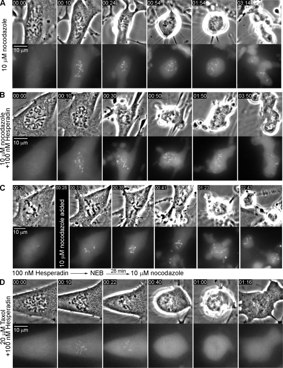
To determine why the duration of mitosis (DM) is less in Taxol than in nocodazole or Eg5 inhibitors we studied the relationship between Taxol concentration, the DM, and the mitotic checkpoint. We found that unlike for other spindle poisons, in Taxol the DM becomes progressively shorter as the concentration surpasses approximately 0.5 microM. Studies on RPE1 and PtK2 expressing GFP/cyclin B or YFP/Mad2 revealed that cells ultimately satisfy the checkpoint in Taxol and do so faster at concentrations >0.5 microM. Inhibiting the aurora-B kinase in Taxol-treated RPE1 cells accelerates checkpoint satisfaction by stabilizing syntelic kinetochore attachments and reduces the DM to approximately 1.5 h regardless of drug concentration. A similar stabilization of syntelic attachments by Taxol itself appears responsible for accelerated checkpoint satisfaction at concentrations >0.5 microM. Our results provide a novel conceptual framework for how Taxol prolongs mitosis and caution against using it in checkpoint studies. They also offer an explanation for why some cells are more sensitive to lower versus higher Taxol concentrations.


Figures

Figure 1.
Figure 1.
The duration of mitosis increases with Taxol concentration until a point after which it decreases. (A–F) Duration of mitosis vs. drug concentration for RPE1 cells treated with nocodazole, Taxol, or Epothilone B, and for PtK2, human BJ fibroblasts, and HeLa cells treated with Taxol. (G) 5 µM Taxol does not shorten mitosis in RPE1 if microtubule assembly is first prevented by vinblastine.
Figure 2.
Figure 2.
In Taxol, Mad2 is progressively depleted from kinetochores until the checkpoint is satisfied (see also Fig. S1). (A–C) YFP/Mad2-PtK2 cells were followed without treatment (A) or in 0.5 (B) or 20 µM (C) Taxol. Top rows are phase-contrast images and bottom rows are maximum intensity projected YFP fluorescence images of the same cell. Exit from mitosis occurs only after the last Mad2-positive kinetochore (A and C, arrowhead) loses its Mad2. (B) In 0.5 µM Taxol a kinetochore (arrowhead) can lose and then reacquire Mad2, as shown in high-magnification images from the boxed regions. Membrane blebbing is typical as cells exit mitosis in Taxol (C) and is not due to apoptosis. (D) Time vs. average number of Mad2-positive kinetochores in controls and after treatment with 10 µM nocodazole or 20 µM Taxol. (E–G) Similar plots of individual controls (E), or cells treated with 0.5 µM (F) or 20 µM Taxol (G). The number of Mad2-positive kinetochores often transiently increases in 0.5 µM but not 20 µM Taxol.
Figure 3.
Figure 3.
Cyclin B/GFP fluorescence decay increases sharply just before Taxol-treated RPE1 cells exit mitosis. (A) In controls, cyclin B/GFP fluorescence exhibits a sudden, sharp, continuous drop several minutes before chromatid disjunction or cytokinesis (D, arrow), which is clear from the time vs. cyclin B/GFP intensity plot (D). A similar sudden sharp drop in cyclin B/GFP fluorescence is also seen shortly before cells treated with 10 µM Taxol (B) or 10 µM Taxol and 100 nM Hesperadin (C) exit mitosis. (D) Typical normalized cyclin B/GFP fluorescence intensity vs. time plots of controls or cells treated with 3.2 µM nocodazole, 0.5, 5, or 10 µM Taxol, or 10 µM Taxol and Hesperadin. Plots of cyclin B decay in 0.5 µM Taxol were difficult to interpret. Time is in h:min.
Figure 4.
Figure 4.
Chromosomes acquire monotelic, merotelic, or syntelic attachments to asters during spindle assembly in Taxol. (A) 12 serial sections through a RPE1 cell from a culture treated for 5 h with 0.5 µM Taxol before fixation and staining for kinetochores (Hec1) and MTs (α-tubulin). Although some kinetochore pairs have acquired amphitelic attachments to adjacent asters (sections 7 and 12, bottom box), others exhibit monotelic (section 16, right-hand box), merotelic (section 13, box), or syntelic (section 7, top box) attachments. (B–D) Inhibiting aurora B does not diminish Mad2 accumulation on kinetochores of nocodazole-treated cells. RPE1 cells were treated for 10 h with 3.2 µM nocodazole (B) or 3.2 µM nocodazole and 100 nM Hesperadin (C) before fixation and staining for centromeres (ACA) and Mad2. (D) The kinetochore-Mad2 staining intensity was found by the Student's t-test to be the same for both conditions. (E–G) When aurora B is inhibited, Mad2 is progressively depleted from kinetochores in Taxol-treated cells. RPE1cultures were incubated in Taxol (0.5 µM or 10 µM) and Hesperadin for 3–8 h and 10 µM MG132 was added 2 h before fixation and staining for centromeres and Mad2. Although most cells exhibited variable amounts of kinetochore-Mad2 (E), others could readily be located under both conditions which had no kinetochore-Mad2 staining (F and G). Arrowheads in F and G note clear syntelic chromosomes.
Figure 5.
Figure 5.
Inhibiting aurora B with Hesperadin does not prevent Mad2 accumulation at kinetochores in nocodazole-treated Mad2/PtK2 cells. (A and B) Mad2 rapidly reappears on kinetochores when Hesperadin-treated cells in metaphase are exposed to nocodazole (C). (D) By contrast, although Mad2 also accumulates at kinetochores as Taxol-treated cells enter mitosis in Hesperadin, it is depleted with time until all kinetochores lack Mad2 after which the cell exits mitosis (see also Fig. 2, C and D).

References

    1. Ahonen L.J., Kallio M.J., Daum J.R., Bolton M., Manke I.A., Yaffe M.B., Stukenberg P.T., Gorbsky G.J. 2005. Polo-like kinase 1 creates the tension-sensing 3F3/2 phosphoepitope and modulates the association of spindle-checkpoint proteins at kinetochores.Curr. Biol. 15:1078–1089 - PubMed
    1. Brinkley B.R., Stubblefield E., Hsu T.C. 1967. The effects of colcemid inhibition and reversal on the fine structure of the mitotic apparatus of Chinese hamster cells in vitro.J. Ultrastruct. Res. 19:1–18 - PubMed
    1. Brito D.A., Rieder C.L. 2006. Mitotic checkpoint slippage in humans occurs via cyclin B destruction in the presence of an active checkpoint.Curr. Biol. 16:1194–1200 - PMC - PubMed
    1. Brito D.A., Rieder C.L. 2009. The ability to survive mitosis in the presence of microtubule poisons differs significantly between human nontransformed (RPE-1) and cancer (U2OS, HeLa) cells.Cell Motil. Cytoskeleton. 66:437–447 - PMC - PubMed
    1. Brito D.A., Yang Z., Rieder C.L. 2008. Microtubules do not promote mitotic slippage when the spindle assembly checkpoint cannot be satisfied.J. Cell Biol. 182:623–629 - PMC - PubMed

Publication types

MeSH terms