. 2009 Sep 15;106(37):15867-72.
doi: 10.1073/pnas.0908032106. Epub 2009 Aug 31.

A broad screen for targets of immune complexes decorating arthritic joints highlights deposition of nucleosomes in rheumatoid arthritis

Affiliations

A broad screen for targets of immune complexes decorating arthritic joints highlights deposition of nucleosomes in rheumatoid arthritis

Paul A Monach et al. Proc Natl Acad Sci U S A. .

Abstract

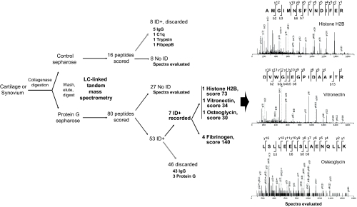
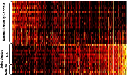
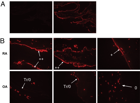
Deposits of Ig and complement are abundant in affected joints of patients with rheumatoid arthritis (RA) and in animal models of RA in which antibodies are demonstrably pathogenic. To identify molecular targets of the Igs deposited in arthritic joints, which may activate local inflammation, we used a combination of mass spectrometry (MS) and protein microarrays. Immune complexes were affinity-purified from surgically removed joint tissues of 26 RA and osteoarthritis (OA) patients. Proteins complexed with IgG were identified by proteomic analysis using tandem MS. A striking diversity of components of the extracellular matrix, and some intracellular components, copurified specifically with IgG from RA and OA tissues. A smaller set of autoantigens was observed only in RA eluates. In complementary experiments, IgG fractions purified from joint immune complexes were tested on protein microarrays against a range of candidate autoantigens. These Igs bound a diverse subset of proteins and peptides from synovium and cartilage, different from that bound by normal serum Ig. One type of intracellular protein detected specifically in RA joints (histones H2A/B) was validated by immunohistology and found to be deposited on the cartilage surface of RA but not OA joints. Thus, autoantibodies to many determinants (whether deposited as "neoantigens" or normal constituents of the extracellular matrix) have the potential to contribute to arthritic inflammation.


Conflict of interest statement

The authors declare no conflict of interest.

Figures

Fig. 1.
Fig. 1.
A typical example of MS analysis of a synovial extract. Synovium from a RA patient was surgically excised, digested with collagenase, and the supernatant divided in half to incubate with proteinG Sepharose or control Sepharose. After extensive washing of the beads, bound material was eluted, reduced and digested exhaustively with trypsin, and loaded onto a microcapillary HPLC system linked to a Q-TOF tandem MS. The remaining peptides detected in the mixtures were analyzed for mass, fragmented, and the ions resulting from fragmentation detected. Data on the masses of each tryptic peptide and of its fragmentation ions were compared to those predicted for all tryptic peptides from all proteins in the NCBInr datase. Among peptides showing reasonable similarity to those in the database (80 in the proteinG eluate and 16 in the control eluate), approximately half were derived from IgG or predictable contaminants, such as keratin, proteinG, or trypsin (ID+, discarded), and many additional peptides did not closely match any known human or human pathogen sequence (no ID). Seven peptides (ID+, recorded) from the proteinG eluate-matched human sequences: 4 derived from fibrinogen and 1 each from histone H2B, vitronectin, and osteoglycin. Visual inspection of the fragmentation spectra for the latter 3 peptides confirmed the assignments, as most masses corresponded closely (delta <0.02) to masses predicted to be formed by the predominant mode of fragmentation (“b” and especially “y” series).
Fig. 2.
Fig. 2.
Binding of joint-eluted IgG vs. normal serum Ig to antigens on a microarray. IgG purified from 16 individual joint extracts from RA and non-RA patients was evaluated for binding to 503 antigens (listed in Table S3) in a microarray format as in Materials and Methods. Comparison was made with 22 normal sera. Colors denote amount of binding: undetectable (black); progressively greater binding (red, yellow, white); full range of scale represents approximately a 1,000-fold difference. Complete numerical data are shown in Dataset S1. Antigens are ordered by difference (fold change) between mean binding by joint-eluted vs. control Ig. Increased binding to 55 antigens was apparent at a level of P < 10−5.
Fig. 3.
Fig. 3.
Immunofluorescence staining of human cartilage samples with a mAb recognizing a complex of H2A/H2B/DNA. Frozen sections were prepared and stained as in Materials and Methods. (A) Staining of a RA cartilage sample with an isotype control mAb (Left) or anti-H2A/H2B/DNA (Right) is shown (10× objective) followed by Texas-red-conjugated goat anti-mouse-IgG. Staining with this control mAb was done in parallel for all samples and was undetectable in all cases. (B) Three examples each of RA (Top) and OA (Bottom) cartilage samples stained with anti-H2A/H2B/DNA are shown (20× objective). Arrows denote the cartilage surface and mAb staining is apparent in red. Examples include the full range of intensities seen, using qualitative descriptions (++, +, Tr/0, 0) as summarized in Table S4. Additional punctate staining seen in all samples represents staining of chondrocyte nuclei, which served as an internal technical positive control. All images have been modified equally, to offset the dimming effect of shrinking them.

Similar articles

Cited by

References

    1. Vander Cruyssen B, et al. Anti-citrullinated protein/peptide antibodies (ACPA) in rheumatoid arthritis: specificity and relation with rheumatoid factor. Autoimmun Rev. 2005;4:468–474. - PubMed
    1. Edwards JC, et al. Efficacy of B-cell-targeted therapy with rituximab in patients with rheumatoid arthritis. N Engl J Med. 2004;350:2572–2581. - PubMed
    1. Monach PA, Benoist C, Mathis D. The role of antibodies in mouse models of rheumatoid arthritis, and relevance to human disease. Adv Immunol. 2004;82:217–248. - PubMed
    1. Kuhn KA, et al. Antibodies against citrullinated proteins enhance tissue injury in experimental autoimmune arthritis. J Clin Invest. 2006;116:961–973. - PMC - PubMed
    1. Ronnelid J, Lysholm J, Engstrom-Laurent A, Klareskog L, Heyman B. Local anti-type II collagen antibody production in rheumatoid arthritis synovial fluid. Evidence for an HLA-DR4-restricted IgG response. Arthritis Rheum. 1994;37:1023–1029. - PubMed

Publication types

MeSH terms