. 2009 Sep 15;106(37):15768-73.
doi: 10.1073/pnas.0906894106. Epub 2009 Aug 31.

Generation of pluripotent stem cells from patients with type 1 diabetes

Affiliations

Generation of pluripotent stem cells from patients with type 1 diabetes

René Maehr et al. Proc Natl Acad Sci U S A. .

Abstract

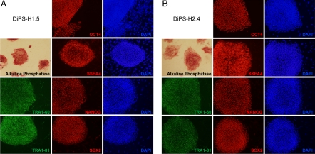
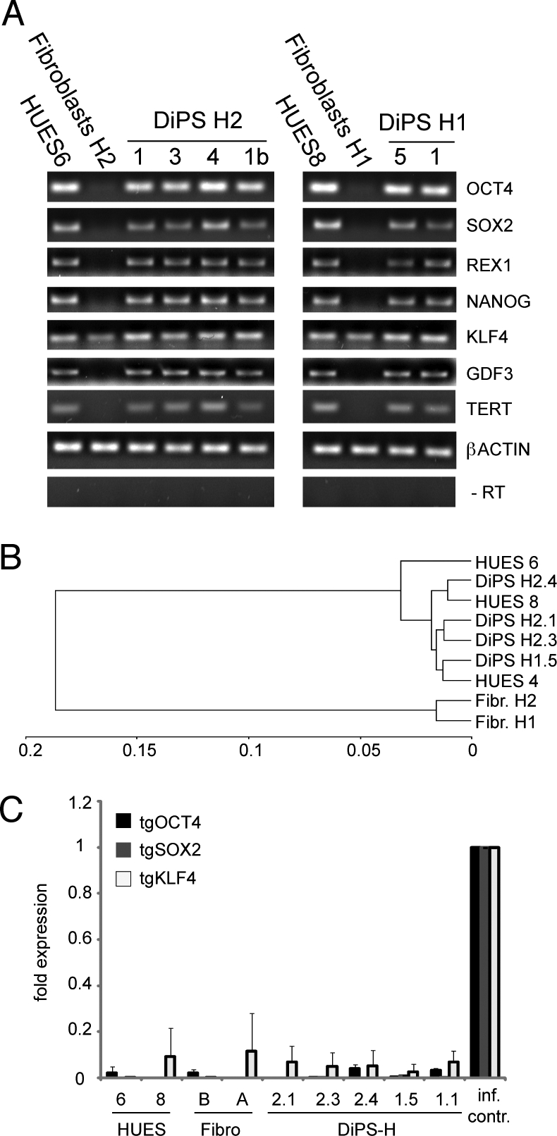
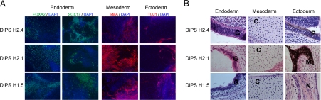
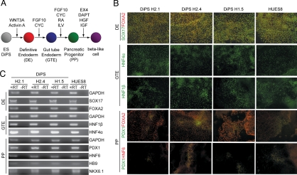
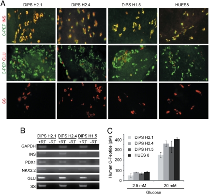
Type 1 diabetes (T1D) is the result of an autoimmune destruction of pancreatic beta cells. The cellular and molecular defects that cause the disease remain unknown. Pluripotent cells generated from patients with T1D would be useful for disease modeling. We show here that induced pluripotent stem (iPS) cells can be generated from patients with T1D by reprogramming their adult fibroblasts with three transcription factors (OCT4, SOX2, KLF4). T1D-specific iPS cells, termed DiPS cells, have the hallmarks of pluripotency and can be differentiated into insulin-producing cells. These results are a step toward using DiPS cells in T1D disease modeling, as well as for cell replacement therapy.


Conflict of interest statement

The authors declare no conflict of interest.

Figures

Fig. 1.
Fig. 1.
Generation of DiPS cells from T1D patients. DiPS lines were established from two T1D affected patient fibroblasts lines H1 (A) and H2 (B). Displayed are DiPS lines (A) H1.5 and (B) H2.4. Detection of AP activity and immunofluorescence analyses for presence of pluripotency markers OCT4, SSEA4, NANOG, TRA1-60, SOX2, and TRA1-81 are indicated. For immunofluorescence stains corresponding nuclear stains (DAPI) visualize all cells including mouse embryonic fibroblast feeder cells.
Fig. 2.
Fig. 2.
Expression analysis of patient specific DiPS cells. (A) Semiquantitative analysis of expression of OCT4, SOX2, REX1, NANOG, KLF4, GDF3, and β ACTIN. Control PCR (no RT) is included. (B) Hierarchical cluster analysis of different DiPS, HUES and fibroblast lines. (C) Quantitative assessment of viral transgene expression (tgOCT4, tgSOX2, and tgKLF4) levels. Viral transgene expression was normalized to control infected BJ fibroblasts (isolation occurred 7 days post infection). Uninfected HUES and fibroblast lines were used as controls. The experiment was performed in duplicates and the error bars represent SD.
Fig. 3.
Fig. 3.
Spontaneous differentiation of DiPS cells into cells of different germ layer origin. (A) In vitro differentiation of DiPS lines H1.5, H2.1, and H2.4 in EB assays was followed by monolayer culture and immunostaining for markers of ectoderm (TUJ1), mesoderm (SMA), and endoderm (FOXA2 and SOX17). An overlay with a nuclear stain (DAPI) is displayed. (B) Teratoma formation occurred after injection of DiPS into immunocompromised mice. Hematoxylin and Eosin staining of teratoma sections shows nerve fibers (N), melanocytes (M), pigmented epithelium (P), cartilage (C), and glandular structures (G).
Fig. 4.
Fig. 4.
Stepwise differentiation of ES/DiPS cells toward β-like cells. (A) Schematic representation of stepwise differentiation of human PS cells to β-like cells. DiPS cell lines H1.5, H2.1, and H2.4 differentiation to definitive endoderm (DE), gut tube endoderm (GTE) and pancreatic progenitors (PPs) indicated by (B) immunostaining and (C) RT-PCR. SOX, SRY (sex determining region Y)-box; FOXA2, forkhead box protein A2; HNF, hepatocyte nuclear factor; PDX1, pancreatic and duodenal homeobox 1; HB9, homeobox gene HLXB9; NKX6.1, NK6 transcription factor related, locus 1.
Fig. 5.
Fig. 5.
DiPS cell lines H1.5, H2.1, and H2.4 differentiate to hormone-expressing endocrine cells indicated by (A) immunostaining and (B) semiquantitative PCR. (C) The DiPS-derived C-peptide-expressing cells secreted C-peptide on glucose stimulation. The DiPS-derived populations were stimulated with 2.5 and 20 mM D-glucose, and the amount of human C-peptide released to culture supernatant was analyzed by ELISA. C-PEP, C-peptide; INS, insulin; GLU, glucagon; SS, somatostatin.

Comment in

  • Stem cells and a cure for type 1 diabetes?
    Todd JA. Todd JA. Proc Natl Acad Sci U S A. 2009 Sep 15;106(37):15523-4. doi: 10.1073/pnas.0908373106. Epub 2009 Sep 9. Proc Natl Acad Sci U S A. 2009. PMID: 19805208 Free PMC article. No abstract available.

Similar articles

Cited by

References

    1. Roep BO. Are insights gained from NOD mice sufficient to guide clinical translation? Another inconvenient truth. Ann N Y Acad Sci. 2007;1103:1–10. - PubMed
    1. Roep BO, Atkinson M. Animal models have little to teach us about type 1 diabetes: 1. In support of this proposal. Diabetologia. 2004;47:1650–1656. - PubMed
    1. von Herrath M, Nepom GT. Animal models of human type 1 diabetes. Nat Immunol. 2009;10:129–132. - PubMed
    1. Dimos JT, et al. Induced Pluripotent Stem Cells Generated from Patients with ALS Can Be Differentiated into Motor Neurons. Science. 2008;321:1218–1221. - PubMed
    1. Ebert AD, et al. Induced pluripotent stem cells from a spinal muscular atrophy patient. Nature. 2009;457:277–280. - PMC - PubMed

Publication types

MeSH terms