. 2009 Dec;8(12):2687-99.
doi: 10.1074/mcp.M900176-MCP200. Epub 2009 Aug 31.

Identification of cardiac myosin-binding protein C as a candidate biomarker of myocardial infarction by proteomics analysis

Affiliations

Identification of cardiac myosin-binding protein C as a candidate biomarker of myocardial infarction by proteomics analysis

Sebastien Jacquet et al. Mol Cell Proteomics. 2009 Dec.

Abstract

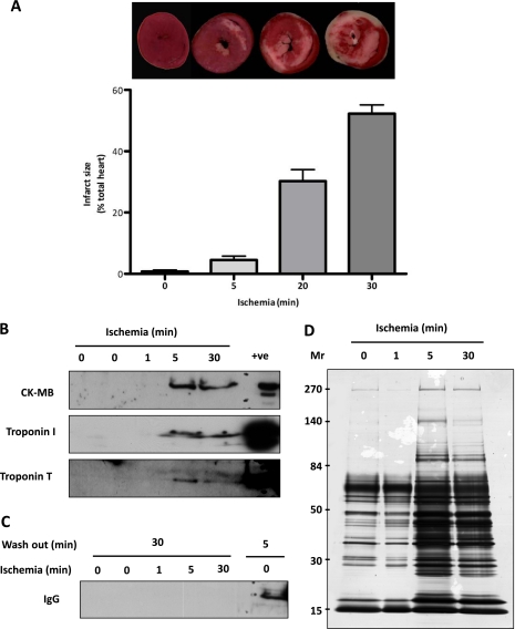
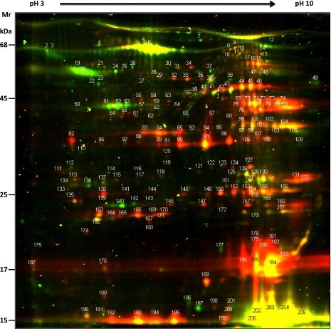
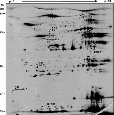
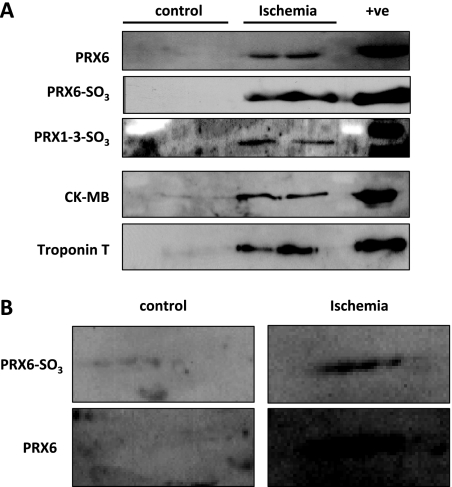
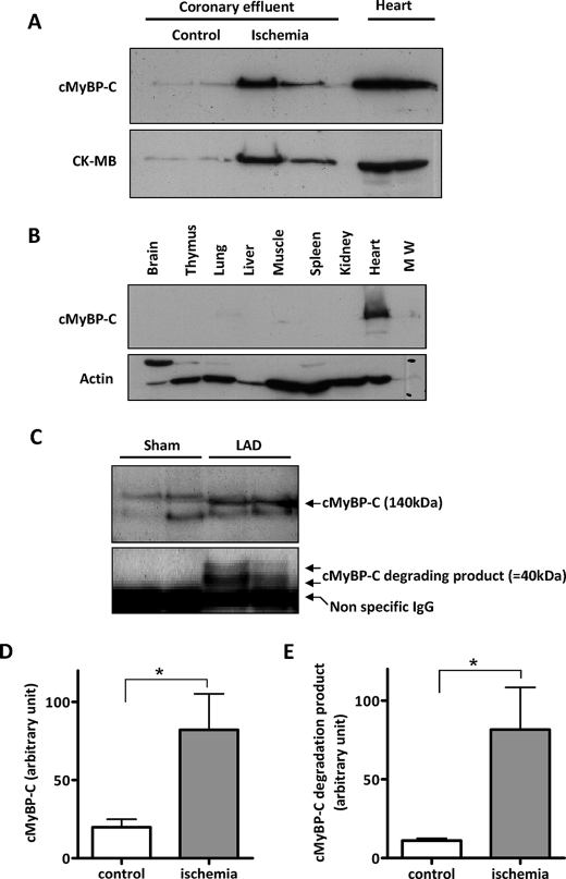
Acute myocardial infarction (AMI) is a common cause of death for which effective treatments are available provided that diagnosis is rapid. The current diagnostic gold standards are circulating cardiac troponins I and T. However, their slow release delays diagnosis, and their persistence limits their utility in the identification of reinfarction. The aim was to identify candidate biomarkers of AMI. Isolated mouse hearts were perfused with oxygenated protein-free buffer, and coronary effluent was collected after ischemia or during matched normoxic perfusion. Effluents were analyzed using proteomics approaches based on one- or two-dimensional initial separation. Of the 459 proteins identified after ischemia with one-dimensional separation, 320 were not detected in the control coronary effluent. Among these were all classic existing biomarkers of AMI. We also identified the cardiac isoform of myosin-binding protein C in its full-length form and as a 40-kDa degradation product. This protein was not detected in the other murine organs examined, increased markedly with even trivial myocardial infarction, and could be detected in the plasma after myocardial infarction in vivo, a profile compatible with a biomarker of AMI. Two-dimensional fluorescence DIGE of ischemic and control coronary effluents identified more than 200 asymmetric spots verified by swapping dyes. Once again existing biomarkers of injury were confirmed as well as posttranslational modifications of antioxidant proteins such as peroxiredoxins. Perfusing hearts with protein-free buffers provides a platform of graded ischemic injury that allows detailed analysis of protein release and identification of candidate cardiac biomarkers like myosin-binding protein C.


Figures

Fig. 1.
Fig. 1.
Characterization of myocardial infarction and coronary effluent protein content. A, relationship between the extent of infarction and duration of ischemia. Isolated retrogradely perfused mouse hearts were subjected to 0, 5, 20, or 30 min of global ischemia (no flow) followed by 2 h of reperfusion. Infarct size was demarcated by TTC staining. Infarct size expressed as mean ± S.E. B, coronary effluent analysis by immunoblotting. Coronary effluents were collected after the indicated duration of global ischemia and probed for CK-MB, troponin I, and troponin T. C, samples were probed for the presence of IgG after 30 or 5 min of perfusion (washout). D, estimation of coronary effluent protein content by silver staining. Coronary effluents (n = 2 per time point) collected at the onset of reperfusion after the indicated period of ischemia were concentrated and analyzed by a 1D gel (5–20%) and silver stained. All bands were excised, and after in gel tryptic digestion, peptides were identified by MS-MS (proteins are listed in supplemental Table 1). +ve, positive control.
Fig. 2.
Fig. 2.
Representative DIGE 2D gel of proteins appearing in coronary effluents after 5 min of global ischemia (Cy5; red) and matched control perfusion (Cy3; green). Coronary effluents from control and ischemic hearts (n = 3–4) were concentrated, and the same amount of protein was CyDye-labeled before separation by 2D gel electrophoresis. The annotated spots were excised and identified by LC-MS/MS (see supplemental Table 2 for the complete list of proteins).
Fig. 3.
Fig. 3.
Representative silver-stained DIGE 2D gel of proteins appearing in coronary effluents after 5 min of global ischemia and matched control perfusion. The known biomarkers identified by mass spectrometry are indicated.
Fig. 4.
Fig. 4.
Verification by immunoblotting of oxidative stress-related proteins appearing in coronary effluent after ischemia. A, analyses of coronary effluents collected after 5 min of ischemia or matching control perfusion by 1D electrophoresis. Peroxiredoxins and their posttranslationally modified oxidized forms (PRX-SO3) were detected only after ischemia. Immunoblots for CK and troponin were used as controls to indicate ischemia-selective protein release. B, analyses of coronary effluents collected after 5 min of ischemia or matching control perfusion by 2D electrophoresis. Immunoblots of a 2D gel (18-cm strip, pH 4–7, 12–20% gradient gel) for peroxiredoxin 6 total and oxidized forms are shown. Peroxiredoxin 6 is present only in ischemic coronary effluent, and the charge train of the oxidized peroxiredoxin-SO3 can be visualized. +ve, positive.
Fig. 5.
Fig. 5.
Proteins released into coronary effluent. Protein release after 5 min of ischemia (identified on the 1D gel) is plotted as -fold change (unique peptide in ischemia/unique peptide in control ratio) on the ordinate against absolute abundance on the abscissa. Abundance is estimated by the obtained spectral count normalized to the molecular weight of the individual protein. The known biomarkers of myocardial infarction are highlighted in red, and proteins in the coronary effluent with a -fold change greater than 5 are numbered and listed in Table IV. MLC, myosin light chain. Asterisk highlights MyBP-C.
Fig. 6.
Fig. 6.
Verification and validation of cMyBP-C as potential new biomarker of acute myocardial infarction. A, 1D separation and immunoblotting of proteins in coronary effluent. Coronary effluents were collected after 5 min of ischemia or matching control perfusion. B, cMyBP-C content among mouse organs. The anti-cMyBP-C antibody reacted only with cardiac tissue. Anti-actin was used as loading control. C, detection of cMyBP-C in the plasma of mice subjected to acute myocardial infarction. Mice were subjected to temporary ligation of the left anterior descending (LAD) coronary artery (30 min) followed by reperfusion (2 h). Blood was collected, and plasma was immunoblotted for cMyBP-C. The full-length cMyBP-C at 140 kDa and the degradation product at 40 kDa were both detected selectively in plasma of mice subjected to temporary, but not sham, coronary artery ligation. D and E, quantification of full-length and short forms of cMyBP-C. Data are derived from n = 6 mice per group and are expressed as mean ± S.E. *, p < 0.05.

References

    1. Rajappa M., Sharma A. ( 2005) Biomarkers of cardiac injury: an update. Angiology 56, 677– 691 - PubMed
    1. Apple F. S., Wu A. H., Mair J., Ravkilde J., Panteghini M., Tate J., Pagani F., Christenson R. H., Mockel M., Danne O., Jaffe A. S. ( 2005) Future biomarkers for detection of ischemia and risk stratification in acute coronary syndrome. Clin. Chem 51, 810– 824 - PubMed
    1. Saenger A. K., Jaffe A. S. ( 2008) Requiem for a heavyweight: the demise of creatine kinase-MB. Circulation 118, 2200– 2206 - PubMed
    1. Pelsers M. M., Hermens W. T., Glatz J. F. ( 2005) Fatty acid-binding proteins as plasma markers of tissue injury. Clin. Chim. Acta 352, 15– 35 - PubMed
    1. Wunderlich M. T., Hanhoff T., Goertler M., Spener F., Glatz J. F., Wallesch C. W., Pelsers M. M. ( 2005) Release of brain-type and heart-type fatty acid-binding proteins in serum after acute ischaemic stroke. J. Neurol 252, 718– 724 - PubMed

Publication types

MeSH terms