Review
. 2009 Sep;73(3):451-80, Table of Contents.
doi: 10.1128/MMBR.00012-09.

The remarkable frequency of human immunodeficiency virus type 1 genetic recombination

Affiliations
Review

The remarkable frequency of human immunodeficiency virus type 1 genetic recombination

Adewunmi Onafuwa-Nuga et al. Microbiol Mol Biol Rev. 2009 Sep.

Abstract

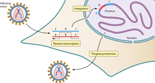
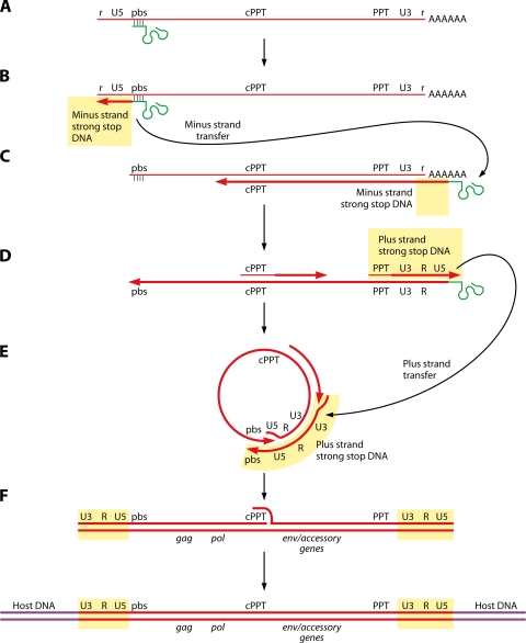
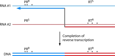
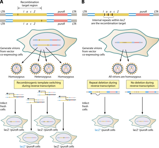
The genetic diversity of human immunodeficiency virus type 1 (HIV-1) results from a combination of point mutations and genetic recombination, and rates of both processes are unusually high. This review focuses on the mechanisms and outcomes of HIV-1 genetic recombination and on the parameters that make recombination so remarkably frequent. Experimental work has demonstrated that the process that leads to recombination--a copy choice mechanism involving the migration of reverse transcriptase between viral RNA templates--occurs several times on average during every round of HIV-1 DNA synthesis. Key biological factors that lead to high recombination rates for all retroviruses are the recombination-prone nature of their reverse transcription machinery and their pseudodiploid RNA genomes. However, HIV-1 genes recombine even more frequently than do those of many other retroviruses. This reflects the way in which HIV-1 selects genomic RNAs for coencapsidation as well as cell-to-cell transmission properties that lead to unusually frequent associations between distinct viral genotypes. HIV-1 faces strong and changeable selective conditions during replication within patients. The mode of HIV-1 persistence as integrated proviruses and strong selection for defective proviruses in vivo provide conditions for archiving alleles, which can be resuscitated years after initial provirus establishment. Recombination can facilitate drug resistance and may allow superinfecting HIV-1 strains to evade preexisting immune responses, thus adding to challenges in vaccine development. These properties converge to provide HIV-1 with the means, motive, and opportunity to recombine its genetic material at an unprecedented high rate and to allow genetic recombination to serve as one of the highest barriers to HIV-1 eradication.


Figures

FIG. 1.
FIG. 1.
Pseudodiploid nature of retroviral virions. Virions that package two genetically distinct copies of viral gRNA (red and blue lines) have the potential to generate recombinant proviruses if one or more recombinogenic template switches (indicated with a dashed black line) occur during reverse transcription. Despite copackaging of two gRNAs, retroviruses are not truly diploid because only one allele at each locus is preserved in the integrated provirus, and any progeny produced from a cell harboring a single recombinant provirus will transmit only one allele at each locus in its progeny.
FIG. 2.
FIG. 2.
Process of HIV-1 reverse transcription. (A) A tRNA primer (tRNAlys) is bound to complementary sequences at the primer binding site (pbs) on encapsidated HIV-1 gRNAs. (B) Minus-strand DNA synthesis initiates from primer tRNA and halts when it reaches the 5′ end of gRNA, generating “minus-strand strong-stop DNA.” (C) After unmasking of the nascent minus-strand strong-stop DNA by RNase H degradation of the template RNA strand, the replicative template switch known as minus-strand transfer occurs for complementary R-region sequences near the 3′ end of the gRNA, and minus-strand DNA synthesis continues. (D) As minus-strand synthesis proceeds, plus-strand DNA synthesis is initiated from oligoribonucleotides that persist at the PPTs. The nascent plus-strand DNA that results when synthesis is halted at the first modified base in primer tRNA is called plus-strand strong-stop DNA. (E) After the RNase H-mediated removal of the tRNA primer, plus-strand strong-stop strand transfer results from the annealing of the 3′ end of plus-strand strong-stop DNA to complementary sequences at the end of the minus-strand DNA intermediate. (F) DNA synthesis is completed, generating double-stranded DNA with long terminal repeats and a central flap at the central PPT (cPPT). Integrase catalyzes the establishment of the provirus, and host repair enzymes remove flaps and gaps. Thin red lines represent viral RNA, and thicker lines represent viral DNA. Primer tRNA is depicted as the green objects. (Data are not to scale.)
FIG. 3.
FIG. 3.
Recombinogenic template switching. The thin lines (blue and red) represent genetically distinct copackaged gRNAs; thick lines represent viral DNA. The arrow shows the direction of DNA synthesis, and asterisks depict the sites of mutations that confer resistance to either RT or PR inhibitors. Here, template switching generates a recombinant provirus that is resistant to both types of inhibitors.
FIG. 4.
FIG. 4.
Retroviral recombination assays. (A) Two vector assay to assess interstrand template switching rates. Each vector has a defect in LacZ-coding sequences, as indicated with the daggers. Each virion generated from producer cells will contain either a homodimer or a heterodimer of gRNAs. Upon infection of fresh cells, recombination between gRNAs within the recombination target region can create a functional lacZ gene for virions containing heterodimeric gRNAs. (B) Single-vector assay demonstrating intrastrand recombination by repeat deletion. The vector contains a repeated sequence within the LacZ coding region. In this instance, virions generated from the producer cell all contain gRNA homodimers. Upon infection, functional lacZ results from a precise deletion of one of the repeats via an intrastrand template switch. Note that repeat deletion can occur either between repeats on one gRNA or from one gRNA to the other: reciprocal repeat assays have shown that intrastrand and interstrand crossovers occur at similar frequencies (240). LTR, long terminal repeat.
FIG. 5.
FIG. 5.
Interactions of HIV-1 RT with primer-templates. HIV-1 RT is represented by the gold oval, where P indicates the DNA polymerase active site and H is the RNase H active site. The thinner line represents the viral RNA template strand, while the thicker line represents the nascent DNA (primer) strand. The blue arrow shows the direction of DNA synthesis. (A) The DNA polymerase active site is engaged at the primer strand 3′ terminus, and the RNase H active site engages the template strand and performs limited template cleavage as DNA synthesis proceeds. (B) When RT reaches the 5′ end of the RNA template, RNase H cleavage leaves an 18-base oligoribonucleotide annealed to the nascent DNA. (C) Upon RT translocation, which displaces the primer terminus from the DNA polymerase active site, or RT rebinding, further RNase H cleavage generates an 8- to 10-base RNA remnant.
FIG. 6.
FIG. 6.
Three models for retroviral recombination. (A) Strand displacement-assimilation showing recombination during plus-strand synthesis. (see the text and reference 160). (B) Forced copy choice (see the text and reference 68). (C) Minus-strand exchange (see the text and reference 26). Thin lines are retroviral RNAs with breaks in the strands. Thick lines represent the nascent DNA; arrows indicate the direction of DNA synthesis. Gold ovals represent HIV-1 RT.
FIG. 7.
FIG. 7.
Speculative model for the processive RT elongation complex. (Based on findings reported in references , , , , and .) (A) Ordinarily, during elongation by RT, RNase H-mediated template strand nicking provides an opportunity for NC protein-facilitated docking of the acceptor template strand onto the nascent DNA primer strand, behind the elongating RT. (B) As the elongating RT continues DNA synthesis on the donor template, secondary RT molecules further degrade template strands, and branch migration brings the acceptor template in proximity to the elongating RT: this is the putative processive RT elongation complex. (C) When RT reaches an impediment to elongation, RNase H activity “catches up” with the elongating RT. (D) Unable to continue DNA synthesis on the donor template, the polymerase active site disengages, and RT translocates as it does at template ends (Fig. 5C), reducing the length of the RNA-DNA hybrid. (E) Further branch migration displaces the residual oligoribonucleotide and forces primer strand realignment onto the homologous portion of the copackaged gRNA, allowing DNA synthesis to proceed, and the eventual completion of an intact provirus. Gold ovals represent HIV-1 RT molecules that contribute to DNA synthesis; green ovals represent secondary RTs that contribute polymerase-independent RNase H activity; circles represent NC (stoichiometry and locations are highly speculative); triangles represent damaged template positions.
FIG. 8.
FIG. 8.
Requirements for generating recombinant genomes. (A) Coinfection with two genetically distinct viruses does not yield recombinants. However, a producer cell must be coinfected with two genetically distinct viruses (shown here as viral particles with two blue or two red RNAs) to produce viral particles with heterodimeric gRNAs. (B) Recombination is observable in cells infected with heterodimeric virions (particle containing one red and one blue RNA strand). Template switching during reverse transcription can generate a recombinant provirus.
FIG. 9.
FIG. 9.
RNA secondary structure in the packaging and dimerization region of HIV-1 gRNA. A region near the 5′ end of the genome, termed Ψ, is responsible for the selective recruitment of gRNA and is coincident with elements required for dimerization between copackaged RNAs. Here, the area is enlarged to show stem-loop 1 (SL1) to stem-loop 4. The gag start codon is indicated as shaded residues; note that alternate models for the 5′ end of HIV-1 gRNA do not include stem-loop 4 but instead evoke long-distance interactions between sequences encompassing the gag start codon and U5 regions (1). Stem-loop 1 is capped by a palindromic sequence known as the DIS, which forms a putative kissing-loop interaction that initiates dimerization between two copackaged gRNAs. (Roles and models for these regions are reviewed in references , , and .)
FIG. 10.
FIG. 10.
Differences between gammaretroviruses and HIV-1 in gRNA selectivity. Shown is a schematic illustration of a single virion-producing cell coinfected with two distinct proviruses, shown as the double-stranded red and blue lines embedded within purple host DNA. Fates of gRNAs during HIV-1 assembly are indicated on the left, and those for the gammaretrovirus MLV are indicated on the right. MLV gRNAs preferentially self-associate at transcription sites in the nucleus, resulting in virions containing mostly homodimerized gRNAs. For HIV-1, RNAs dimerize at random, presumably in the cytoplasm, generating virions with a Hardy-Weinberg distribution of homodimers and heterodimers.
FIG. 11.
FIG. 11.
Retroviral homologous and nonhomologous recombination. (A) Homologous recombination during HIV-1 DNA synthesis. Template switching occurs from one RNA to colinear identical sequences on the second RNA strand. (B to D) Microhomology-guided nonhomologous recombination during reverse transcription. (B) Template switching to a position more 5′ on the acceptor template to generate a deletion. (C) Template switching to a position more 3′ on the acceptor template to generate a duplication. (D) Insertion in deletion, or ectopic duplication, generated by template switching to the acceptor at a more 3′ position, continued synthesis, and a second template switch back to the donor strand at a more 5′ position. The arrows show the direction of DNA synthesis on two copackaged gRNAs. Shaded boxed sequences are regions of homology between donor and acceptor templates.
FIG. 12.
FIG. 12.
Model for host oncogene transduction by animal retroviruses. After a rare chance event leads to the integration of a provirus DNA upstream of a cellular oncogene, readthrough of viral polyadenylation signal transcription can result in the “capture” of downstream oncogene sequences near the 3′ end of a readthrough RNA. Following packaging into a virion and infection of a fresh cell, this readthrough RNA can serve as a nonhomologous recombination template during reverse transcription. In most cases, oncogene transduction results in a replication-defective virus (307).
FIG. 13.
FIG. 13.
Augmentation of HIV-1 multidrug resistance via human sequence transduction. Shown is a schematic overview of the mechanism by which an HIV-1 strain gained enhanced resistance to multiple RT inhibitors during replication in a Japanese child. (A) The top line shows a preinsertion sequence of the HIV-1 strain replicating in the boy's parents and in the child prior to the acquisition of drug resistance; the lower line indicates the sequence of the multidrug-resistant isolate. Blue shading indicates the region of a 30/31 match to human chromosome (chr.) 17. (B) Proposed mechanism for the acquisition of human sequences. (i) Establishment of a provirus upstream of the transduced sequences and generation of a readthrough RNA. (ii) Nonhomologous recombination between viral and human sequences. (iii) Further point mutagenesis during replication in the patient to generate the sequences observed for the patient (314).
FIG. 14.
FIG. 14.
Candidate host sequence insertions in lentivirus genomes. (A) HIV-1 AVT codon-rich env variable region insertions. (Based on patient env sequence data as analyzed by in reference .) The x axis indicates boundaries of genetic segments within the gp120-encoding portions of env. Blue bands indicate proportions of all codons within each genetic region that are AVT (where V is A,C, or G) codons for Asn, Thr, and Ser; orange bands indicate proportions of all codons that are other Asn, Thr, and Ser codons. Note that the most prevalent AVT codon observed in HIV-1 variable regions, AAT, is also the most common trinucleotide repeat in human microsatellite DNA (306). (B) Length variation that emerged in macaques infected with an SIV molecular clone (Based on data presented in reference .) Shown are V1 region amino acid sequences for the parental Mne strain (GenBank accession number M32741) and the nine variant classes reported after the development of AIDS-like symptoms (accession numbers M79283, M79284, M79287, M79288, M79286, M79289, M79285, M79299, and M79293). Nucleotide sequences were used in unfiltered blastN to query all macaque sequences in GenBank. As indicated by the expect values (E values are similar to P values) presented at the right, length variation in all but one variant (variant 3) significantly increased the similarity of that SIV strain to macaque sequences under stringent search parameters (+1/−4 match/mismatch penalties), suggesting that these inserts, which lack significant similarity to the parental Mne genome, were host derived. (C) Host-like insertion between sequences derived from two different subtypes in a new HIV-1 CRF. (Based on data presented in reference .) The figure indicates the genome positions where bootstrap analyses indicated breakpoints between subtypes in CRF15_01B. The dotted circle and lines and aligned sequences at the bottom indicate the location of a region of 30 bases of identity between this HIV-1 isolate (GenBank accession number AF530576) and human chromosome 3 (accession number NT_005612.15), which is assigned an expect value of 0.001 in unfiltered blastN with default search parameters. Note that this structure is reminiscent of the host-derived inserts that frequently splint experimentally derived nonhomologous crossovers (87).

References

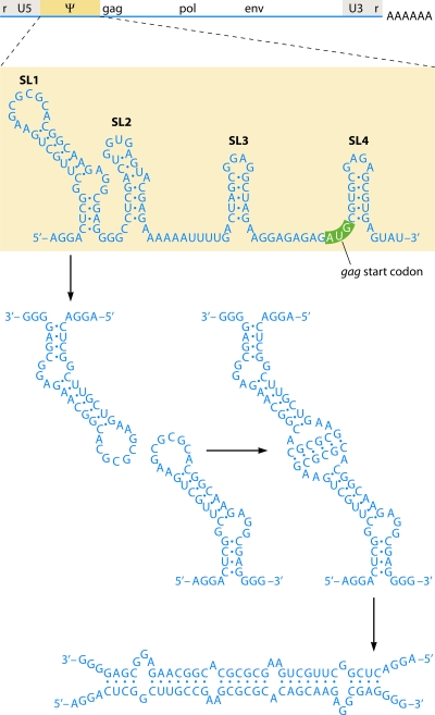
    1. Abbink, T. E., and B. Berkhout. 2003. A novel long distance base-pairing interaction in human immunodeficiency virus type 1 RNA occludes the Gag start codon. J. Biol. Chem. 27811601-11611. - PubMed
    1. Abbondanzieri, E. A., G. Bokinsky, J. W. Rausch, J. X. Zhang, S. F. Le Grice, and X. Zhuang. 2008. Dynamic binding orientations direct activity of HIV reverse transcriptase. Nature 453184-189. - PMC - PubMed
    1. Abecasis, A. B., P. Lemey, N. Vidal, T. de Oliveira, M. Peeters, R. Camacho, B. Shapiro, A. Rambaut, and A. M. Vandamme. 2007. Recombination confounds the early evolutionary history of human immunodeficiency virus type 1: subtype G is a circulating recombinant form. J. Virol. 818543-8551. - PMC - PubMed
    1. Allen, T. M., and M. Altfeld. 2003. HIV-1 superinfection. J. Allergy Clin. Immunol. 112829-835. - PubMed
    1. Allen, T. M., M. Altfeld, S. C. Geer, E. T. Kalife, C. Moore, K. M. O'Sullivan, I. Desouza, M. E. Feeney, R. L. Eldridge, E. L. Maier, D. E. Kaufmann, M. P. Lahaie, L. Reyor, G. Tanzi, M. N. Johnston, C. Brander, R. Draenert, J. K. Rockstroh, H. Jessen, E. S. Rosenberg, S. A. Mallal, and B. D. Walker. 2005. Selective escape from CD8+ T-cell responses represents a major driving force of human immunodeficiency virus type 1 (HIV-1) sequence diversity and reveals constraints on HIV-1 evolution. J. Virol. 7913239-13249. - PMC - PubMed

Publication types