

Review
. 2009 Sep;73(3):542-63, Table of Contents.
doi: 10.1128/MMBR.00009-09.
Lessons in signaling and tumorigenesis from polyomavirus middle T antigen
Affiliations
- PMID: 19721090
- PMCID: PMC2738132
- DOI: 10.1128/MMBR.00009-09
Item in Clipboard
Review
Lessons in signaling and tumorigenesis from polyomavirus middle T antigen
Microbiol Mol Biol Rev.
2009 Sep.
Abstract
The small DNA tumor viruses have provided a very long-lived source of insights into many aspects of the life cycle of eukaryotic cells. In recent years, the emphasis has been on cancer-related signaling. Here we review murine polyomavirus middle T antigen, its mechanisms, and its downstream pathways of transformation. We concentrate on the MMTV-PyMT transgenic mouse, one of the most studied models of breast cancer, which permits the examination of in situ tumor progression from hyperplasia to metastasis.
Figures
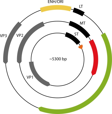
The polyomavirus genome. Early transcription of the T antigens proceeds in a clockwise direction. Late transcription of the capsid proteins (VP1, VP2, and VP3) is counterclockwise. Replication and transcription are controlled by the enhancer (ENH) and the origin of viral DNA replication (ORI). T-antigen coding sequences are in different colors to emphasize reading frame differences.
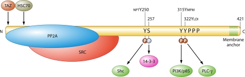
MT and its direct interactions with cellular proteins. SRC, Src family tyrosine kinase (Src, Yes, Fyn). The binding proteins Shc, PI3K, phosphoinositide 3-kinase, and PLCγ1, which bind phosphotyrosine sequences, are shown in dark green. The sequences near the tyrosine phosphorylation sites are shown above the sequence. 14-3-3, which binds pSer257, is shown in magenta. PPP represents the proline rich region. The light green area represents the hydrophobic membrane attachment site. The C-terminal sequence is AHSM384QRHLRRLGR393TLLLVTFLAALLGICLMLFILI KRSRHF421 (underlining indicates the hydrophobic stretch). Proteins such as Grb2 that associate with MT partners are not shown here.
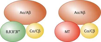
PP2A and MT-PP2A complexes. For PP2A, two different A subunit scaffolds, α and β, bind one of two different catalytic C subunits and one of very many different B family regulatory subunits. MT replaces B subunits in the complex.
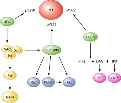
Simplified diagram of signaling pathways downstream of MT phosphotyrosines.
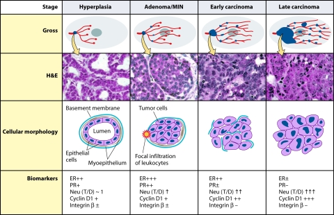
Summary of tumor progression and biomarker expression in the MMTV-PyMT634 mouse model of breast cancer. Four tumor stages are observed in transgenic MMTV-PyMT634 mice at different ages and in ducts that are at different stages of development. At the earliest stage (2 to 4 weeks after birth), growth of the short, underdeveloped prepubertal ducts takes place from the bulbous end buds under the influence of growth factors. In sexually mature mice (from 6 weeks on, when adenomas are observed), ducts continue to grow from the end buds under the control of estrogen and growth factors until they reach the confines of the gland. (The figure and legend [see below] are adapted from reference with permission from the American Society for Investigative Pathology.) The Gross panel displays the overall development of lesions in mammary glands of PyMT mice. Tumor lesions are indicated by blue dots. The hematoxylin-and-eosin panel displays the corresponding histology of primary lesions at different stages of tumor progression. The cellular morphology panel schematically illustrates changes in the cytology of the cells as well as the integrity of the basement membrane and the presence or absence of myoepithelial and focal inflammation. Moreover, the changes in biomarkers during tumor progression are summarized in the panel of biomarkers. T/D, the ratio of Neu expression between lesions and normal ducts in age-matched mammary glands.
References
-
- Aguzzi, A., E. F. Wagner, R. L. Williams, and S. A. Courtneidge. 1990. Sympathetic hyperplasia and neuroblastomas in transgenic mice expressing polyoma middle T antigen. New Biol. 2533-543. - PubMed
-
- Allison, A. C., J. N. Monga, and V. Hammond. 1974. Increased susceptibility to virus oncogenesis of congenitally thymus-deprived nude mice. Nature 252746-747. - PubMed
-
- Almholt, K., A. Juncker-Jensen, O. D. Laerum, K. Dano, M. Johnsen, L. R. Lund, and J. Romer. 2008. Metastasis is strongly reduced by the matrix metalloproteinase inhibitor Galardin in the MMTV-PymT transgenic breast cancer model. Mol. Cancer Ther. 72758-2767. - PubMed
Publication types
MeSH terms
Substances
Grants and funding
LinkOut - more resources
Full Text Sources
Other Literature Sources
