. 2009 Mar;19(2):117-25.
doi: 10.1055/s-0028-1096209.

Imaging findings of rhinocerebral mucormycosis

Affiliations

Imaging findings of rhinocerebral mucormycosis

Diego A Herrera et al. Skull Base. 2009 Mar.

Abstract

Background and objectives: The purpose of this study was to describe common radiographic patterns that may be useful in predicting the diagnosis of rhinocerebral mucormycosis.

Methods: We retrospectively evaluated the imaging and clinical data of four males and one female, 3 to 72 years old, with rhinocerebral mucormycosis.

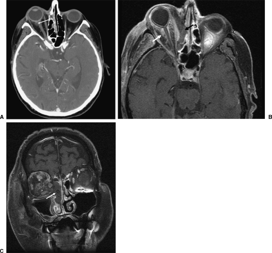
Results: All the patients presented with sinusitis and ophthalmological symptoms. Most of the patients (80%) had isointense lesions relative to brain in T1-weighted images. The signal intensity in T2-weighted images was more variable, with only one (20%) patient showing hyperintensity. A pattern of anatomic involvement affecting the nasal cavity, maxillary sinus, orbit, and ethmoid cells was consistently observed in all five patients (100%). Our series demonstrated a mortality rate of 60%.

Conclusion: Progressive and rapid involvement of the cavernous sinus, vascular structures and intracranial contents is the usual evolution of rhinocerebral mucormycosis. In the context of immunosupression, a pattern of nasal cavity, maxillary sinus, ethmoid cells, and orbit inflammatory lesions should prompt the diagnosis of mucormycosis. Multiplanar magnetic resonance imaging shows anatomic involvement, helping in surgery planning. However, the prognosis is grave despite radical surgery and antifungals.

Keywords: MRI; Rhinocerebral mucormycosis; imaging findings; neuroradiology.


Figures

Figure 1
Figure 1
A 72-year-old male diabetic patient presented with right eye pain and proptosis. (A) Computed tomography axial image shows subtle increased density in intraconal fat in the right orbit (arrow), representing early inflammatory process. (B, C) T1 fat-saturated images obtained after the administration of gadolinium the same day demonstrate progression of infection, with orbital invasion and severe compromise of left nasal cavity and ethmoid cells (arrows). Fast interval change is suggestive of rhinocerebral mucormycosis.
Figure 2
Figure 2
A 72-year-old male diabetic patient presented with right eye pain and proptosis. (A) Magnetic resonance imaging (MRI) control after right orbital exenteration. Despite amphotericin treatment and surgical debriding, cavernous sinus compromise and abnormal signal in contact with the carotid wall (arrow) was noted after 3 months. (B) Interval progression with carotid wall invasion (arrow) was noted during follow up. (C) Catheter angiography shows linear and irregular filling of the cavernous portion of the internal carotid artery (arrow) confirming MRI findings. Vascular invasion is a frequent feature of mucormycosis infection.
Figure 3
Figure 3
A 16-year-old female patient with leukemia in remission presented with facial and ocular pain. Computed tomography coronal image shows an inflammatory process involving nasal cavity, ethmoid cells, maxillary sinus, and orbit in the left side (arrows). Combined involvement of the previously mentioned anatomic structures should raise suspicion of the diagnosis of invasive fungal sinusitis.
Figure 4
Figure 4
A 16-year-old female patient with leukemia in remission presented with facial and ocular pain, status post right orbital exenteration and debriding. (A) Magnetic resonance imaging fat-saturated post gadolinium T1 coronal image shows an inflammatory lesion involving the cribriform plate (arrow). (B) Interval change is noted, with increasing thickness of dural enhancement (arrow), corresponding to intracranial extension, which is a frequent finding in mucormycosis infection.
Figure 5
Figure 5
A 45-year-old diabetic patient presented with orbital symptoms and fifth cranial nerve palsy. (A) Magnetic resonance imaging (MRI) fat-saturated T1 post gadolinium coronal image shows lack of enhancement of the left cavernous sinus (arrow), corresponding to mucormycosis invasion. (B) MRI post gadolinium T1-weighted axial image shows perineural spread through the left fifth cranial nerve with brainstem and middle cerebellar peduncle invasion (arrows). (C) MR diffusion-weighted image shows restricted diffusion (arrow). (D) MR fat-saturated T1 post gadolinium coronal image shows interval progression in invasion of the cavernous sinus and carotid artery (arrow). Perineural (V3) spread through the foramen ovale is also noted (arrowhead). (E) MR angiography time of flight shows thinning of the left cavernous internal carotid artery and MCA branches (arrows). (F) MR flair axial image shows left hemispheric infarcts (arrows).
Figure 6
Figure 6
A 13-year-old male patient with blastic anemia and neutropenia. (A) Computed tomography coronal image shows an extensive inflammatory process involving the nasal cavity, ethmoid cells, maxillary, frontal sinus, and orbit in the left, which may raise the suspicion of invasive fungal infection (arrows). Facial soft tissues are also involved (arrowheads). (B) Magnetic resonance fat-saturated T1-weighted axial image shows pial/arachnoid enhancement corresponding to leptomeningeal mucormycosis spread (arrows). Soft tissue compromise is again noted (arrowheads).
Figure 7
Figure 7
Intraoperative photo of hard palate prior to maxillectomy, taken ~6 hours after presentation. On initial examination this lesion (arrow) was a subcentimeter area of pallor on the left, at the junction between the hard and soft palates.
Figure 8
Figure 8
Intraoperative photo of left maxillectomy and orbital exenteration, inferior view. Note the extent of necrosis of the palate (arrow).
Figure 9
Figure 9
Histologic sections of periorbital tissue with multiple, broad nonseptate hyphae (arrows) surrounded by inflammatory infiltrate (hematoxylin & eosin, 400 ×).

References

    1. Rumboldt Z, Castillo M. Indolent intracranial mucormycosis: case report. AJNR Am J Neuroradiol. 2002;23:932–934. - PMC - PubMed
    1. Chan L L, Singh S, Jones D, et al. Imaging of mucormycosis skull base osteomyelitis. AJNR Am J Neuroradiol. 2000;21:828–831. - PMC - PubMed
    1. Anselmo-Lima W T, Lopes R P, Valera F C, et al. Invasive fungal rhinosinusitis in immunocompromised patients. Rhinology. 2004;42:141–144. - PubMed
    1. Paulltauf A. Mycosis mucorina. Virchows Arch. 1885;102:543.
    1. Naussbaum E S, Holl W A. Rhinocerebral mucormycosis: changing patterns of disease. Surg Neurol. 1994;41:152–156. - PubMed

LinkOut - more resources