. 2009 Oct 23;424(1):153-61.
doi: 10.1042/BJ20082377.

Regulation of Rnd3 localization and function by protein kinase C alpha-mediated phosphorylation

Affiliations

Regulation of Rnd3 localization and function by protein kinase C alpha-mediated phosphorylation

James P Madigan et al. Biochem J. .

Abstract

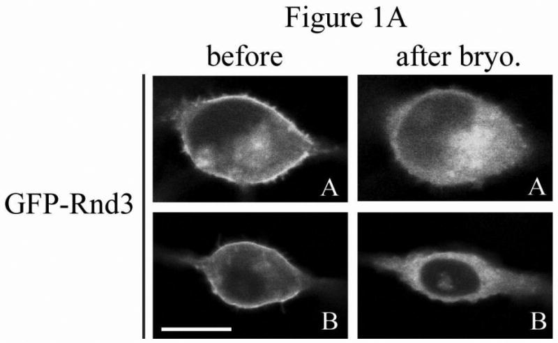
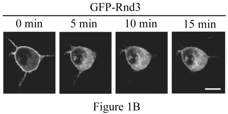
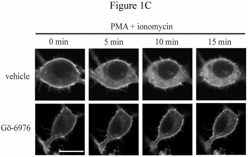
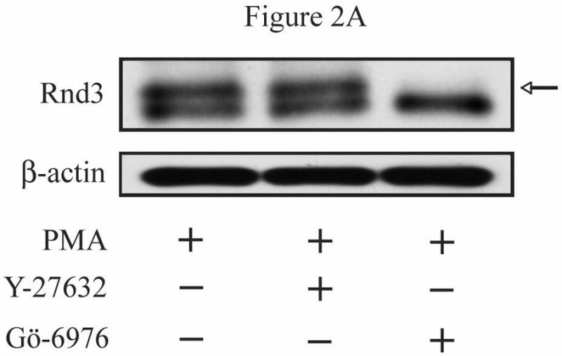
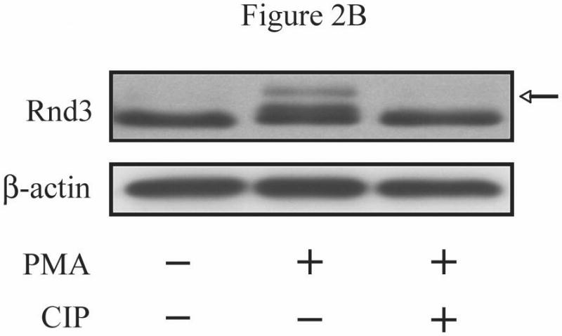
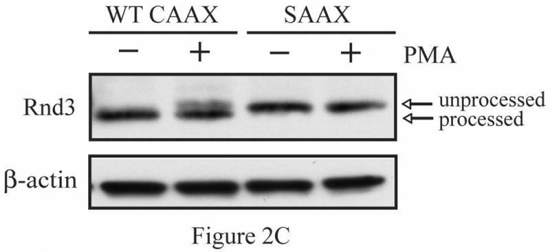
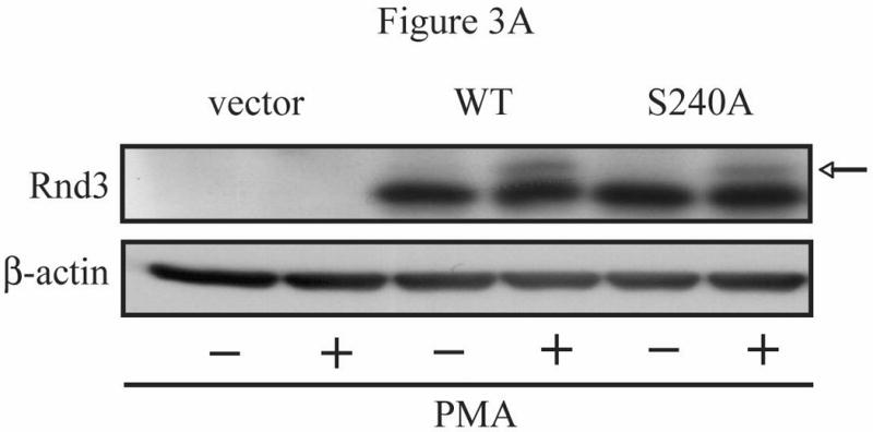
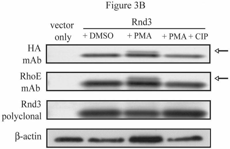
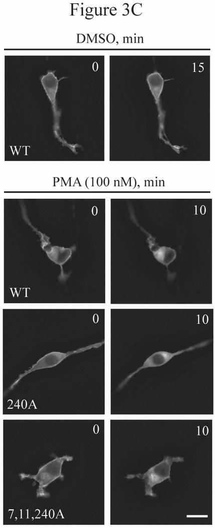
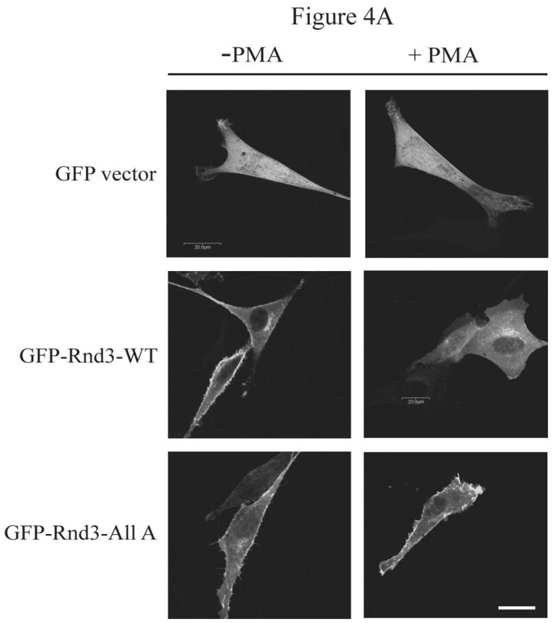
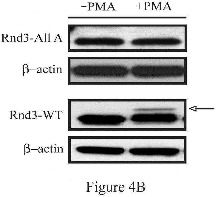
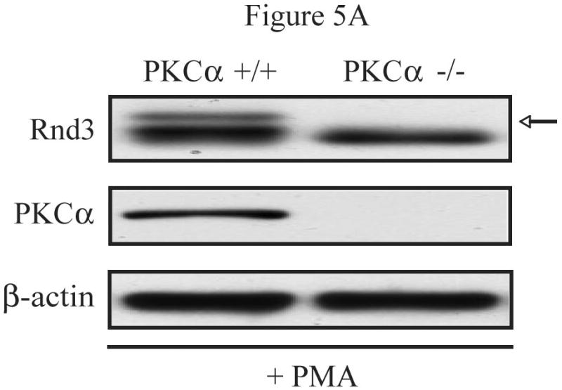
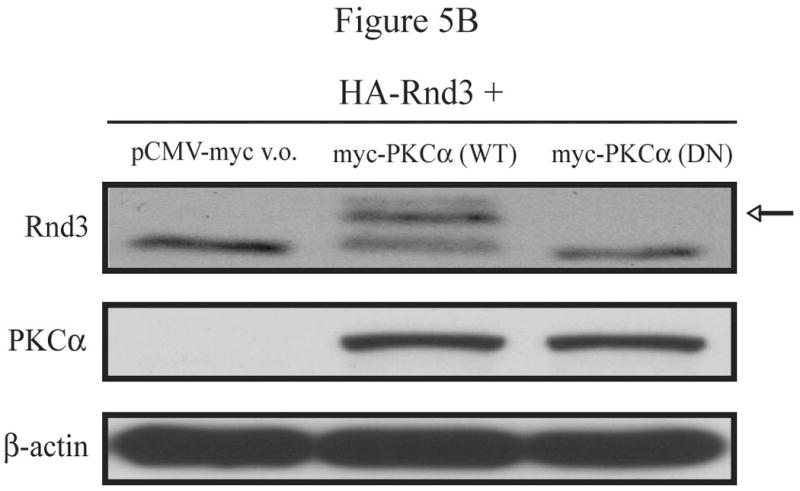
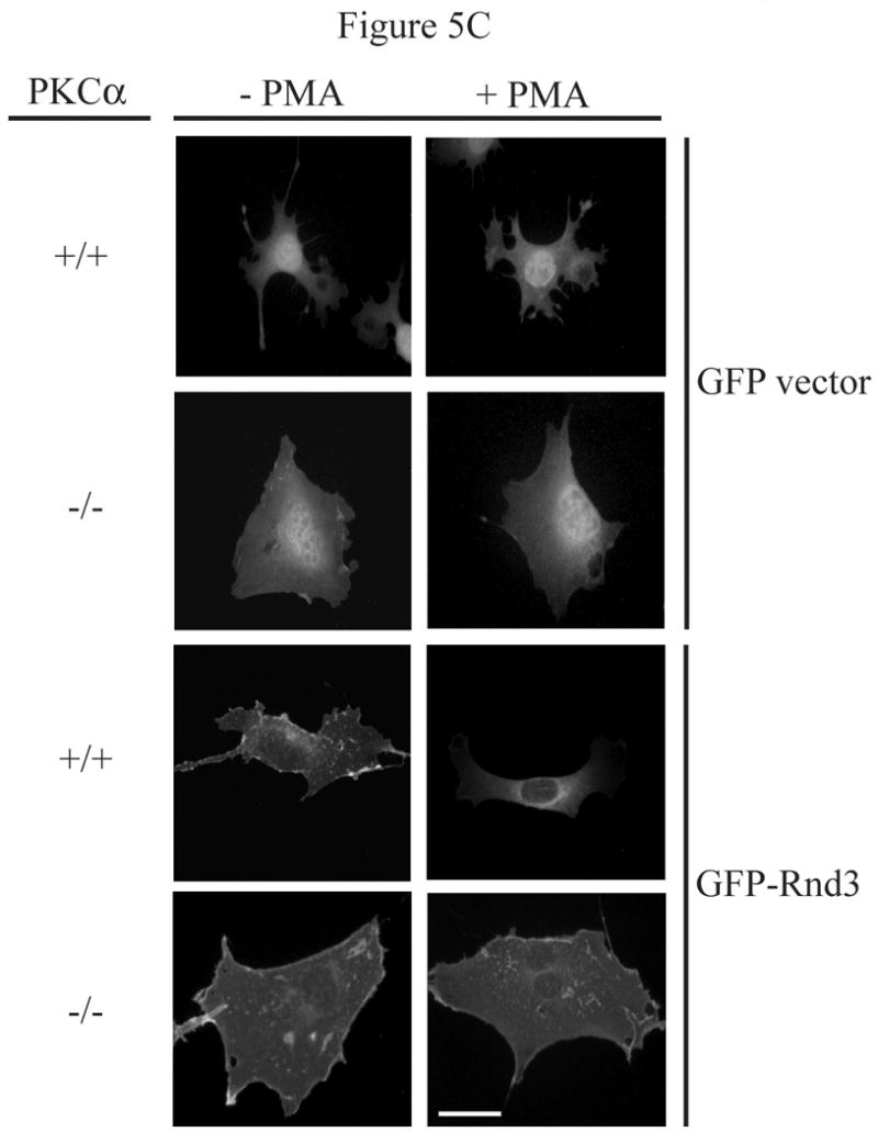
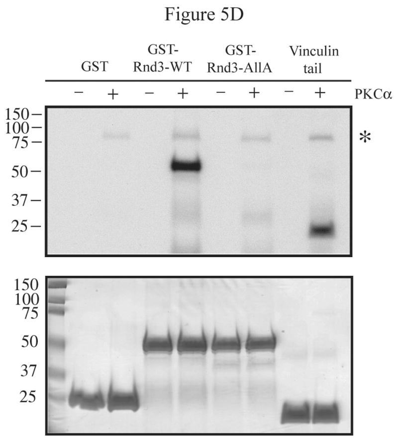
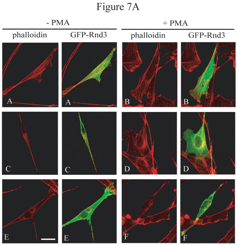
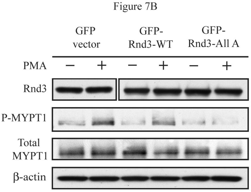
The Rnd proteins (Rnd1, Rnd2 and Rnd3/RhoE) form a distinct branch of the Rho family of small GTPases. Altered Rnd3 expression causes changes in cytoskeletal organization and cell cycle progression. Rnd3 functions to decrease RhoA activity, but how Rnd3 itself is regulated to cause these changes is still under investigation. Unlike other Rho family proteins, Rnd3 is regulated not by GTP/GDP cycling, but at the level of expression and by post-translational modifications such as prenylation and phosphorylation. We show in the present study that, upon PKC (protein kinase C) agonist stimulation, Rnd3 undergoes an electrophoretic mobility shift and its subcellular localization becomes enriched at internal membranes. These changes are blocked by inhibition of conventional PKC isoforms and do not occur in PKCalpha-null cells or to a non-phosphorylatable mutant of Rnd3. We further show that PKCalpha directly phosphorylates Rnd3 in an in vitro kinase assay. Additionally, we provide evidence that the phosphorylation status of Rnd3 has a direct effect on its ability to block signalling from the Rho-ROCK (Rho-kinase) pathway. These results identify an additional mechanism of regulation and provide clarification of how Rnd3 modulates Rho signalling to alter cytoskeletal organization.


Figures

Figure 1
Figure 1
Rnd3 localization is altered after PKC activation. (A) PKC agonist bryostatin-1 causes loss of Rnd3 from the plasma membrane. NIH 3T3 cells transiently expressing GFP-Rnd3 were treated with bryostatin-1 (100 nM). Representative live images of two separate cells before (left panel) and 10 min after (right panel) addition of agonist are shown. (B) Activation of PKC by using PMA + ionomycin also causes loss of Rnd3 from the plasma membrane. NIH 3T3 cells transiently expressing GFP-Rnd3 were treated with PMA (100 nM) + ionomycin (500 μg/mL). Live images are shown of a single cell visualized at 5 min increments. (C) Inhibitor of conventional PKCs blocks Rnd3 translocation. NIH 3T3 cells transiently expressing GFP-Rnd3 were treated with DMSO vehicle or Gö-6976 (2.5 μM) to inhibit conventional PKCs for 3 h prior to stimulation with PMA (100 nM) + ionomycin (500 μg/mL). Live images are shown of single cells visualized at 5 min increments. Scale bar is 20 μm.
Figure 1
Figure 1
Rnd3 localization is altered after PKC activation. (A) PKC agonist bryostatin-1 causes loss of Rnd3 from the plasma membrane. NIH 3T3 cells transiently expressing GFP-Rnd3 were treated with bryostatin-1 (100 nM). Representative live images of two separate cells before (left panel) and 10 min after (right panel) addition of agonist are shown. (B) Activation of PKC by using PMA + ionomycin also causes loss of Rnd3 from the plasma membrane. NIH 3T3 cells transiently expressing GFP-Rnd3 were treated with PMA (100 nM) + ionomycin (500 μg/mL). Live images are shown of a single cell visualized at 5 min increments. (C) Inhibitor of conventional PKCs blocks Rnd3 translocation. NIH 3T3 cells transiently expressing GFP-Rnd3 were treated with DMSO vehicle or Gö-6976 (2.5 μM) to inhibit conventional PKCs for 3 h prior to stimulation with PMA (100 nM) + ionomycin (500 μg/mL). Live images are shown of single cells visualized at 5 min increments. Scale bar is 20 μm.
Figure 2
Figure 2
Activation of conventional PKCs causes a phosphorylation-dependent mobility shift of Rnd3. (A) NIH 3T3 cells expressing HA-Rnd3 were pretreated for 3 h with either DMSO vehicle, Y-27632 (10 μM) or Gö-6976 (2.5 μM). Cells were then treated with PMA (100 nM) for 10 min and cell lysates were resolved on SDS-PAGE. The slower migrating Rnd3 band (arrow) was seen in both the vehicle and the Y-27632 pretreated cells, but not in cells pretreated with the conventional PKC inhibitor Gö-6976. (B) Calf intestinal phosphatase (CIP) treatment causes disappearance of the slower migrating band of Rnd3 (arrow). NIH 3T3 cells transiently expressing HA-Rnd3 expression vector were treated with PMA (100 nM) + ionomycin (500 μg/mL). CIP was added to the cell lysate to reverse phosphorylation. Cell lysates were resolved on SDS-PAGE and blotted with anti-HA antibody. (C) A CAAX mutant of Rnd3 does not shift after activation of PKCs. NIH 3T3 cells expressing HA-tagged WT Rnd3 and a SAAX mutant were treated with PMA (100 nM) for 10 min and cell lysates were resolved on SDS-PAGE.
Figure 2
Figure 2
Activation of conventional PKCs causes a phosphorylation-dependent mobility shift of Rnd3. (A) NIH 3T3 cells expressing HA-Rnd3 were pretreated for 3 h with either DMSO vehicle, Y-27632 (10 μM) or Gö-6976 (2.5 μM). Cells were then treated with PMA (100 nM) for 10 min and cell lysates were resolved on SDS-PAGE. The slower migrating Rnd3 band (arrow) was seen in both the vehicle and the Y-27632 pretreated cells, but not in cells pretreated with the conventional PKC inhibitor Gö-6976. (B) Calf intestinal phosphatase (CIP) treatment causes disappearance of the slower migrating band of Rnd3 (arrow). NIH 3T3 cells transiently expressing HA-Rnd3 expression vector were treated with PMA (100 nM) + ionomycin (500 μg/mL). CIP was added to the cell lysate to reverse phosphorylation. Cell lysates were resolved on SDS-PAGE and blotted with anti-HA antibody. (C) A CAAX mutant of Rnd3 does not shift after activation of PKCs. NIH 3T3 cells expressing HA-tagged WT Rnd3 and a SAAX mutant were treated with PMA (100 nM) for 10 min and cell lysates were resolved on SDS-PAGE.
Figure 3
Figure 3
Multiple residues of Rnd3 are involved in the PKC-dependent translocation and gel mobility shift. (A) The phosphodeficient S240A mutant still displays a mobility shift. NIH 3T3 cells transiently expressing HA-Rnd3 proteins were treated with PMA (100 nM) for 10 min. Cell lysates were resolved on SDS-PAGE and immunoblotted with anti-HA antibody. The phosphodeficient Rnd3 S240A mutant still displayed a mobility shift (arrow), indicating that the shift is not caused by phosphorylation at S240. (B) An N-terminal peptide polyclonal antibody (aa 1-15) does not recognize the mobility-shifted form of Rnd3. NIH 3T3 cells transiently expressing HA-Rnd3 or empty vector were treated with either DMSO vehicle or PMA (100 nM) for 10 min -/+ CIP treatment. Cell lysates were resolved on SDS-PAGE and immunoblotted initially with anti-HA antibody. The blot was then stripped and blotted sequentially with a specific anti-Rnd3/RhoE antibody and anti-Rnd3 antiserum. The lack of a mobility-shifted form of HA-Rnd3 when using the N-terminal Rnd3 peptide polyclonal antibody indicates that the shifted form of Rnd3 (arrow) is dependent on the sequence found in the first 15 residues. (C) GFP-Rnd3 multiple phosphorylation mutants still translocate from the plasma membrane after PKC activation. NIH 3T3 cells were transiently transfected with either GFP-Rnd3-WT or GFP-Rnd3 phosphomutants and treated with either vehicle or PMA (100 nM) for the indicated times. Live images were taken on a confocal microscope. No differences in plasma membrane localization were noted between WT Rnd3 and the Rnd3 phosphorylation mutants after treatment with PMA. Scale bar is 20 μm.
Figure 3
Figure 3
Multiple residues of Rnd3 are involved in the PKC-dependent translocation and gel mobility shift. (A) The phosphodeficient S240A mutant still displays a mobility shift. NIH 3T3 cells transiently expressing HA-Rnd3 proteins were treated with PMA (100 nM) for 10 min. Cell lysates were resolved on SDS-PAGE and immunoblotted with anti-HA antibody. The phosphodeficient Rnd3 S240A mutant still displayed a mobility shift (arrow), indicating that the shift is not caused by phosphorylation at S240. (B) An N-terminal peptide polyclonal antibody (aa 1-15) does not recognize the mobility-shifted form of Rnd3. NIH 3T3 cells transiently expressing HA-Rnd3 or empty vector were treated with either DMSO vehicle or PMA (100 nM) for 10 min -/+ CIP treatment. Cell lysates were resolved on SDS-PAGE and immunoblotted initially with anti-HA antibody. The blot was then stripped and blotted sequentially with a specific anti-Rnd3/RhoE antibody and anti-Rnd3 antiserum. The lack of a mobility-shifted form of HA-Rnd3 when using the N-terminal Rnd3 peptide polyclonal antibody indicates that the shifted form of Rnd3 (arrow) is dependent on the sequence found in the first 15 residues. (C) GFP-Rnd3 multiple phosphorylation mutants still translocate from the plasma membrane after PKC activation. NIH 3T3 cells were transiently transfected with either GFP-Rnd3-WT or GFP-Rnd3 phosphomutants and treated with either vehicle or PMA (100 nM) for the indicated times. Live images were taken on a confocal microscope. No differences in plasma membrane localization were noted between WT Rnd3 and the Rnd3 phosphorylation mutants after treatment with PMA. Scale bar is 20 μm.
Figure 4
Figure 4
GFP-Rnd3-WT, but not GFP-Rnd3-All A, translocate from the plasma membrane after PKC activation. (A) NIH 3T3 cells were transiently transfected with either GFP-vector, GFP-Rnd3-WT or GFP-Rnd3-All A. Cells were treated with either vehicle or PMA (100 nM) for 10 min. Live cell images were taken on a confocal microscope. PMA treatment causes loss of GFP-WT-Rnd3 from the plasma membrane along with a corresponding flattened phenotype. A similar event was not seen in GFP-Rnd3-All A expressing cells. Scale bar is 20 μm. (B) FLAG-Rnd3-WT, but not FLAG-Rnd3-All A, displays a gel mobility shift on SDS-PAGE after PKC activation. NIH 3T3 cells were transiently transfected with either FLAG-Rnd3-WT or FLAG-Rnd3-All A and treated with either vehicle or PMA (100 nM) for 10 min. Lysates were resolved on SDS-PAGE and probed with anti-FLAG antibody to visualize FLAG-tagged Rnd3 protein. Only FLAG-Rnd3-WT was shifted in the presence of PMA (arrow).
Figure 4
Figure 4
GFP-Rnd3-WT, but not GFP-Rnd3-All A, translocate from the plasma membrane after PKC activation. (A) NIH 3T3 cells were transiently transfected with either GFP-vector, GFP-Rnd3-WT or GFP-Rnd3-All A. Cells were treated with either vehicle or PMA (100 nM) for 10 min. Live cell images were taken on a confocal microscope. PMA treatment causes loss of GFP-WT-Rnd3 from the plasma membrane along with a corresponding flattened phenotype. A similar event was not seen in GFP-Rnd3-All A expressing cells. Scale bar is 20 μm. (B) FLAG-Rnd3-WT, but not FLAG-Rnd3-All A, displays a gel mobility shift on SDS-PAGE after PKC activation. NIH 3T3 cells were transiently transfected with either FLAG-Rnd3-WT or FLAG-Rnd3-All A and treated with either vehicle or PMA (100 nM) for 10 min. Lysates were resolved on SDS-PAGE and probed with anti-FLAG antibody to visualize FLAG-tagged Rnd3 protein. Only FLAG-Rnd3-WT was shifted in the presence of PMA (arrow).
Figure 5
Figure 5
PKCα is the isozyme responsible for Rnd3 phosphorylation. (A) PMA stimulation causes a gel mobility-shift of Rnd3 in WT MEF cells, but not in PKCα knock-out MEFs. PKCα knock-out MEFs and matched WT MEFs transiently expressing HA-Rnd3 were treated with PKC agonist PMA (100 nM) for 10 min. Cell lysates were resolved on SDS-PAGE and blotted with anti-HA antibody. Cell lysates were further probed with an anti-PKCα antibody to confirm absence of PKCα protein expression in knock-out MEF cells. The slower migrating band of Rnd3 was seen only in WT MEF cells (arrow). (B) Reintroduction of PKCα-WT, but not of PKCα-DN, into PKCα knock-out MEFS causes a mobility shift of Rnd3 when cells are treated with PKC agonist PMA. PKCα knock-out MEFs transiently expressing HA-Rnd3 along with either pCMV-vector, Myc- PKCα-WT or Myc-PKCα-DN were treated with PKC agonist PMA (100 nM) for 10 min. Cell lysates were resolved on SDS-PAGE and probed with anti-HA antibody. A gel mobility shift of Rnd3 (arrow) was seen only in lysates from PKCα knock-out cells when WT PKCα was reintroduced. (C) GFP-Rnd3 translocates from the plasma membrane in WT MEF cell, but not PKCα knock-out MEFs, after PKC stimulation. WT MEFs and PKCα knock-out MEFs were transiently transfected with either GFP vector or GFP-Rnd3. MEFs were treated with PKC agaonist PMA (100nM) for 10 min and live cell images were taken. Scale bar is 20 μm. (D) Rnd3 is phosphorylated by PKCα in vitro. GST alone, GST-Rnd3-WT, GST-Rnd3-All A and a tail fragment of vinculin (aa 881-1135) were used as substrates in a PKCα in vitro kinase assay. Rnd3-WT, but not Rnd3-All A, incorporates 32P-labelled phosphate. * autophosphorylated PKCα.
Figure 5
Figure 5
PKCα is the isozyme responsible for Rnd3 phosphorylation. (A) PMA stimulation causes a gel mobility-shift of Rnd3 in WT MEF cells, but not in PKCα knock-out MEFs. PKCα knock-out MEFs and matched WT MEFs transiently expressing HA-Rnd3 were treated with PKC agonist PMA (100 nM) for 10 min. Cell lysates were resolved on SDS-PAGE and blotted with anti-HA antibody. Cell lysates were further probed with an anti-PKCα antibody to confirm absence of PKCα protein expression in knock-out MEF cells. The slower migrating band of Rnd3 was seen only in WT MEF cells (arrow). (B) Reintroduction of PKCα-WT, but not of PKCα-DN, into PKCα knock-out MEFS causes a mobility shift of Rnd3 when cells are treated with PKC agonist PMA. PKCα knock-out MEFs transiently expressing HA-Rnd3 along with either pCMV-vector, Myc- PKCα-WT or Myc-PKCα-DN were treated with PKC agonist PMA (100 nM) for 10 min. Cell lysates were resolved on SDS-PAGE and probed with anti-HA antibody. A gel mobility shift of Rnd3 (arrow) was seen only in lysates from PKCα knock-out cells when WT PKCα was reintroduced. (C) GFP-Rnd3 translocates from the plasma membrane in WT MEF cell, but not PKCα knock-out MEFs, after PKC stimulation. WT MEFs and PKCα knock-out MEFs were transiently transfected with either GFP vector or GFP-Rnd3. MEFs were treated with PKC agaonist PMA (100nM) for 10 min and live cell images were taken. Scale bar is 20 μm. (D) Rnd3 is phosphorylated by PKCα in vitro. GST alone, GST-Rnd3-WT, GST-Rnd3-All A and a tail fragment of vinculin (aa 881-1135) were used as substrates in a PKCα in vitro kinase assay. Rnd3-WT, but not Rnd3-All A, incorporates 32P-labelled phosphate. * autophosphorylated PKCα.
Figure 6
Figure 6
PKCα-dependent Rnd3 phosphorylation downregulates Rnd3 inhibitory activity and leads to increased signaling through the Rho-ROCK pathway. (A) Treatment with PMA causes reformation of stress fibers in GFP-Rnd3-WT but not in GFP-Rnd3-All A expressing cells. NIH 3T3 cells were transiently transfected with either GFP-vector (A and B), GFP-Rnd3-WT (C and D) or GFP-Rnd3-All A phosphorylation mutant (E and F) and treated with either DMSO vehicle (A,C,E) or PMA (100 nM) for minutes (B,D,F). Live images were taken on a confocal microscope. Actin structures were visualized with phalloidin. PMA treatment caused loss of GFP-WT-Rnd3 from the plasma membrane along with a corresponding flattened phenotype. Furthermore, there was a reappearance of stress fibers seen in GFP-Rnd3-WT expressing cells when treated with PMA. Similar events were not seen in GFP-Rnd3-All A expressing cells. Scale bar is 20 μm. (B) PKCα-dependent phosphorylation leads to increased signaling through the Rho-ROCK pathway. NIH 3T3 cells transientlty expressing GFP-vector, GFP-Rnd3-WT or GFP-Rnd3-All A were treated similarly as above. Lysates were separated by SDS-PAGE and probed with anti-GFP-antibody to visualize equal expression of the GFP-fusion proteins and GFP protein alone. Additionally, lysates were probed with anti-P-MYPT1 antibody to detect signaling from the Rho-ROCK pathway. An increase in P-MYPT1 signal was seen only in GFP-Rnd3-WT- and not in GFP-Rnd3-All A-expressing cells, indicating increased signaling through the Rho-ROCK pathway only in the presence of phosphorylatable Rnd3.
Figure 6
Figure 6
PKCα-dependent Rnd3 phosphorylation downregulates Rnd3 inhibitory activity and leads to increased signaling through the Rho-ROCK pathway. (A) Treatment with PMA causes reformation of stress fibers in GFP-Rnd3-WT but not in GFP-Rnd3-All A expressing cells. NIH 3T3 cells were transiently transfected with either GFP-vector (A and B), GFP-Rnd3-WT (C and D) or GFP-Rnd3-All A phosphorylation mutant (E and F) and treated with either DMSO vehicle (A,C,E) or PMA (100 nM) for minutes (B,D,F). Live images were taken on a confocal microscope. Actin structures were visualized with phalloidin. PMA treatment caused loss of GFP-WT-Rnd3 from the plasma membrane along with a corresponding flattened phenotype. Furthermore, there was a reappearance of stress fibers seen in GFP-Rnd3-WT expressing cells when treated with PMA. Similar events were not seen in GFP-Rnd3-All A expressing cells. Scale bar is 20 μm. (B) PKCα-dependent phosphorylation leads to increased signaling through the Rho-ROCK pathway. NIH 3T3 cells transientlty expressing GFP-vector, GFP-Rnd3-WT or GFP-Rnd3-All A were treated similarly as above. Lysates were separated by SDS-PAGE and probed with anti-GFP-antibody to visualize equal expression of the GFP-fusion proteins and GFP protein alone. Additionally, lysates were probed with anti-P-MYPT1 antibody to detect signaling from the Rho-ROCK pathway. An increase in P-MYPT1 signal was seen only in GFP-Rnd3-WT- and not in GFP-Rnd3-All A-expressing cells, indicating increased signaling through the Rho-ROCK pathway only in the presence of phosphorylatable Rnd3.

Similar articles

Cited by

References

    1. Hall A. Rho GTPases and the actin cytoskeleton. Science. 1998;279:509–514. - PubMed
    1. Nobes CD, Hall A. Rho, rac and cdc42 GTPases: regulators of actin structures, cell adhesion and motility. Biochem Soc Trans. 1995;23:456–459. - PubMed
    1. Ridley AJ, Hall A. The small GTP-binding protein rho regulates the assembly of focal adhesions and actin stress fibers in response to growth factors. Cell. 1992;70:389–399. - PubMed
    1. Ridley AJ, Paterson HF, Johnston CL, Diekmann D, Hall A. The small GTP-binding protein rac regulates growth factor-induced membrane ruffling. Cell. 1992;70:401–410. - PubMed
    1. Kozma R, Ahmed S, Best A, Lim L. The Ras-related protein Cdc42Hs and bradykinin promote formation of peripheral actin microspikes and filopodia in Swiss 3T3 fibroblasts. Mol Cell Biol. 1995;15:1942–1952. - PMC - PubMed

Publication types