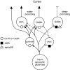
Activation of the basal forebrain by the orexin/hypocretin neurones
- PMID: 19723027
- PMCID: PMC2938067
- DOI: 10.1111/j.1748-1716.2009.02036.x
Activation of the basal forebrain by the orexin/hypocretin neurones
Abstract
The orexin neurones play an essential role in driving arousal and in maintaining normal wakefulness. Lack of orexin neurotransmission produces a chronic state of hypoarousal characterized by excessive sleepiness, frequent transitions between wake and sleep, and episodes of cataplexy. A growing body of research now suggests that the basal forebrain (BF) may be a key site through which the orexin-producing neurones promote arousal. Here we review anatomical, pharmacological and electrophysiological studies on how the orexin neurones may promote arousal by exciting cortically projecting neurones of the BF. Orexin fibres synapse on BF cholinergic neurones and orexin-A is released in the BF during waking. Local application of orexins excites BF cholinergic neurones, induces cortical release of acetylcholine and promotes wakefulness. The orexin neurones also contain and probably co-release the inhibitory neuropeptide dynorphin. We found that orexin-A and dynorphin have specific effects on different classes of BF neurones that project to the cortex. Cholinergic neurones were directly excited by orexin-A, but did not respond to dynorphin. Non-cholinergic BF neurones that project to the cortex seem to comprise at least two populations with some directly excited by orexin-A that may represent wake-active, GABAergic neurones, whereas others did not respond to orexin-A but were inhibited by dynorphin and may be sleep-active, GABAergic neurones. This evidence suggests that the BF is a key site through which orexins activate the cortex and promote behavioural arousal. In addition, orexins and dynorphin may act synergistically in the BF to promote arousal and improve cognitive performance.
Conflict of interest statement
Figures


Similar articles
-
Dynorphin inhibits basal forebrain cholinergic neurons by pre- and postsynaptic mechanisms.J Physiol. 2016 Feb 15;594(4):1069-85. doi: 10.1113/JP271657. Epub 2016 Jan 5. J Physiol. 2016. PMID: 26613645 Free PMC article.
-
Innervation of orexin/hypocretin neurons by GABAergic, glutamatergic or cholinergic basal forebrain terminals evidenced by immunostaining for presynaptic vesicular transporter and postsynaptic scaffolding proteins.J Comp Neurol. 2006 Dec 1;499(4):645-61. doi: 10.1002/cne.21131. J Comp Neurol. 2006. PMID: 17029265 Free PMC article.
-
Opposite effects of noradrenaline and acetylcholine upon hypocretin/orexin versus melanin concentrating hormone neurons in rat hypothalamic slices.Neuroscience. 2005;130(4):807-11. doi: 10.1016/j.neuroscience.2004.10.032. Neuroscience. 2005. PMID: 15652980
-
Experience-dependent plasticity in hypocretin/orexin neurones: re-setting arousal threshold.Acta Physiol (Oxf). 2010 Mar;198(3):251-62. doi: 10.1111/j.1748-1716.2009.02047.x. Epub 2009 Sep 24. Acta Physiol (Oxf). 2010. PMID: 19785627 Free PMC article. Review.
-
Orexin/hypocretin modulation of the basal forebrain cholinergic system: insights from in vivo microdialysis studies.Pharmacol Biochem Behav. 2008 Aug;90(2):156-62. doi: 10.1016/j.pbb.2008.01.008. Epub 2008 Jan 19. Pharmacol Biochem Behav. 2008. PMID: 18281084 Review.
Cited by
-
Descending projections from the basal forebrain to the orexin neurons in mice.J Comp Neurol. 2017 May 1;525(7):1668-1684. doi: 10.1002/cne.24158. Epub 2017 Feb 22. J Comp Neurol. 2017. PMID: 27997037 Free PMC article.
-
Role of adenosine and wake-promoting basal forebrain in insomnia and associated sleep disruptions caused by ethanol dependence.J Neurochem. 2010 Nov;115(3):782-94. doi: 10.1111/j.1471-4159.2010.06980.x. Epub 2010 Sep 28. J Neurochem. 2010. PMID: 20807311 Free PMC article.
-
Orexin A-induced enhancement of attentional processing in rats: role of basal forebrain neurons.Psychopharmacology (Berl). 2016 Feb;233(4):639-47. doi: 10.1007/s00213-015-4139-z. Epub 2015 Nov 4. Psychopharmacology (Berl). 2016. PMID: 26534765 Free PMC article.
-
Promotion of Wakefulness and Energy Expenditure by Orexin-A in the Ventrolateral Preoptic Area.Sleep. 2015 Sep 1;38(9):1361-70. doi: 10.5665/sleep.4970. Sleep. 2015. PMID: 25845696 Free PMC article.
-
Pareidolias: complex visual illusions in dementia with Lewy bodies.Brain. 2012 Aug;135(Pt 8):2458-69. doi: 10.1093/brain/aws126. Epub 2012 May 30. Brain. 2012. PMID: 22649179 Free PMC article.
References
-
- Abrahamson EE, Leak RK, Moore RY. The suprachiasmatic nucleus projects to posterior hypothalamic arousal systems. Neuroreport. 2001;12:435–40. - PubMed
-
- Arrigoni E, Chamberlin NL, Saper CB, McCarley RW. Adenosine inhibits basal forebrain cholinergic and noncholinergic neurons in vitro. Neuroscience. 2006;140:403–13. - PubMed
Publication types
MeSH terms
Substances
Grants and funding
LinkOut - more resources
Full Text Sources
Miscellaneous

