Review
. 2009 Sep 1:7:77.
doi: 10.1186/1479-5876-7-77.

Recent progress towards development of effective systemic chemotherapy for the treatment of malignant brain tumors

Affiliations
Review

Recent progress towards development of effective systemic chemotherapy for the treatment of malignant brain tumors

Hemant Sarin. J Transl Med. .

Abstract

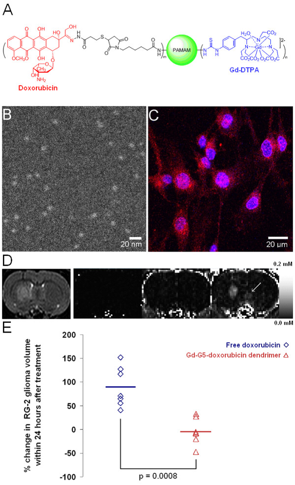
Systemic chemotherapy has been relatively ineffective in the treatment of malignant brain tumors even though systemic chemotherapy drugs are small molecules that can readily extravasate across the porous blood-brain tumor barrier of malignant brain tumor microvasculature. Small molecule systemic chemotherapy drugs maintain peak blood concentrations for only minutes, and therefore, do not accumulate to therapeutic concentrations within individual brain tumor cells. The physiologic upper limit of pore size in the blood-brain tumor barrier of malignant brain tumor microvasculature is approximately 12 nanometers. Spherical nanoparticles ranging between 7 nm and 10 nm in diameter maintain peak blood concentrations for several hours and are sufficiently smaller than the 12 nm physiologic upper limit of pore size in the blood-brain tumor barrier to accumulate to therapeutic concentrations within individual brain tumor cells. Therefore, nanoparticles bearing chemotherapy that are within the 7 to 10 nm size range can be used to deliver therapeutic concentrations of small molecule chemotherapy drugs across the blood-brain tumor barrier into individual brain tumor cells. The initial therapeutic efficacy of the Gd-G5-doxorubicin dendrimer, an imageable nanoparticle bearing chemotherapy within the 7 to 10 nm size range, has been demonstrated in the orthotopic RG-2 rodent malignant glioma model. Herein I discuss this novel strategy to improve the effectiveness of systemic chemotherapy for the treatment of malignant brain tumors and the therapeutic implications thereof.


Figures

Figure 1
Figure 1
Synthesis of gadolinium (Gd)-diethyltriaminepentaacetic acid (DTPA) conjugated polyamidoamine (PAMAM) dendrimers and images of higher generation (G) Gd-dendrimers with annular dark-field scanning transmission electron microscopy. A) Illustrations of naked PAMAM dendrimer generations from the ethylenediamine core (G0) to generation 3 (G3). The exterior of naked PAMAM dendrimers is positively charged due to the presence of terminal amine groups. The number of terminal amine groups doubles with each successive generation. B) Synthetic scheme for the production of Gd-DTPA conjugated PAMAM dendrimers. The conjugation of Gd-DTPA (charge -2) to the terminal amine groups neutralizes the positive charge on the dendrimer exterior. C) Annular dark-field scanning transmission electron microscopy images of Gd-G5, Gd-G6, Gd-G7, and Gd-G8 dendrimers adsorbed onto an ultrathin carbon support film. The average diameter of sixty Gd-G7 dendrimers is 11.0 ± 0.7 nm and that of sixty Gd-G8 dendrimers is 13.3 ± 1.4 nm (mean ± standard deviation). Scale bar = 20 nm. Adapted from reference[73].
Figure 2
Figure 2
Dynamic contrast-enhanced MRI-based Gd concentration maps of Gd-dendrimer distribution within large and small RG-2 rodent gliomas over time. A) Large RG-2 gliomas. Gd-G1 thorough Gd-G7 dendrimers extravasate across the BBTB of the microvasculature of large RG-2 gliomas. After extravasating across the BBTB, Gd-G1 through Gd-G4 dendrimers only remain temporarily within the extravascular compartment of tumor tissue, as these lower Gd-dendrimer generations maintain peak blood concentrations for only a few minutes. The Gd-G5 through Gd-G7 dendrimers accumulate over time within the extravascular compartment of tumor tissue, as these generations maintain peak blood concentrations for several hours. The Gd-G8 dendrimers remain intravascular, since Gd-G8 dendrimers are larger than the physiologic upper limit of pore size in the BBTB of large RG-2 gliomas. RG-2 glioma volumes (mm3): Gd-G1, 104; Gd-G2, 94; Gd-G3, 94; lowly conjugated (LC) Gd-G4, 162; Gd-G4, 200; Gd-G5, 230; Gd-G6, 201; Gd-G7, 170; Gd-G8, 289. B) Small RG-2 gliomas. Gd-G1 thorough Gd-G6 dendrimers extravasate across the BBTB of the microvasculature of small RG-2 gliomas. Since small RG-2 gliomas are less vascular than large RG-2 gliomas, there is a relative lack of accumulation of the lower Gd-dendrimer generations in the extravascular compartment of small RG-2 gliomas as compared to large RG-2 gliomas (panel A). This is especially evident in the case of Gd-G1 dendrimers, which maintain peak blood concentrations for the shortest time period of all the Gd-dendrimer generations. Gd-G5 and Gd-G6 dendrimers accumulate over time within the extravascular compartment of even the small RG-2 gliomas, since these generations maintain peak blood concentrations fro several hours and are smaller than the physiologic upper limit of pore size in the BBTB. Both Gd-G7 and Gd-G8 dendrimers remain intravascular in small RG-2 gliomas, since both Gd-G7 and Gd-G8 dendrimers are larger than the physiologic upper limit of pore size in the BBTB of small RG-2 gliomas. RG-2 glioma volumes (mm3): Gd-G1, 27; Gd-G2, 28; Gd-G3, 19; LC Gd-G4, 24; Gd-G4, 17; Gd-G5, 18; Gd-G6, 22; Gd-G7, 24; Gd-G8, 107. Respective Gd-dendrimer generations administered intravenously over 1 minute at a Gd dose of 0.09 mmol Gd/kg animal body weight. Scale ranges from 0 mM [Gd] to 0.1 mM [Gd]. Adapted from reference[73].
Figure 3
Figure 3
Steady-state blood concentrations of successively higher generation Gd-dendrimers over time in rodents. Gd-G1 dendrimers (MW 6 kDa), Gd-G2 dendrimers (MW 11 kDa), Gd-G3 dendrimers (MW 19 kDa), lowly conjugated (LC) Gd-G4 dendrimers (MW 25 kDa), and standard Gd-G4 dendrimers (MW 40 kDa) maintain peak blood concentrations for only a few minutes. Gd-G5 dendrimers (MW 80 kDa) maintain peak blood concentrations for over 2 hours. Gd-G6 dendrimers (MW 130 kDa), Gd-G7 dendrimers (MW 330 kDa), and Gd-G8 dendrimers (MW 597 kDa) also maintain peak blood concentrations for over 2 hours similar to those of Gd-G5 dendrimers (concentration profiles not shown for purposes of figure clarity). Respective Gd-dendrimer generations administered intravenously over 1 minute at a Gd dose of 0.09 mmol Gd/kg animal body weight. Blood concentrations of Gd-dendrimers over time measured in the superior sagittal sinus. Gd-G1 (n = 4), Gd-G2 (n = 6), Gd-G3 (n = 6), lowly conjugated (LC) Gd-G4 (n = 4), Gd-G4 (n = 6), Gd-G5 (n = 6), Gd-G6 (n = 5), Gd-G7 (n = 5), and Gd-G8 (n = 6). Error bars represent standard deviations. Adapted from reference[73].
Figure 4
Figure 4
Synthesis of rhodamine B dye (RB) conjugated Gd-dendrimers and fluorescence microscopy of rhodamine B conjugated Gd-dendrimer uptake in cultured RG-2 glioma cells versus in RG-2 glioma cells of harvested RG-2 glioma tumor specimens. A) Synthetic scheme for production of rhodamine B dye conjugated Gd-dendrimers. Rhodamine B and DTPA are conjugated to the naked dendrimer terminal amines via stable covalent bonds. In functionalized dendrimers, approximately 35% of the terminal amines are occupied by Gd-DTPA, and approximately 7% of the terminal amines are occupied by rhodamine B. B) In vitro fluorescence microscopy of cultured RG-2 glioma cells incubated for 4 hours in media containing either rhodamine B conjugated Gd-G2 dendrimers (left), rhodamine B conjugated Gd-G5 dendrimers (middle), or rhodamine B conjugated Gd-G8 dendrimers (right) at a concentration of 7.2 μM with respect to rhodamine B. Scale bars = 20 μm. Rhodamine B conjugated Gd-G2 dendrimers enter RG-2 glioma cells, and in some cases, the cell nuclei (left). Rhodamine B conjugated Gd-G5 dendrimers (middle) and rhodamine B conjugated Gd-G8 dendrimers (right) enter the cytoplasm of RG-2 glioma cells, but do not localize within the nuclei. C) Dynamic contrast-enhanced MRI-based Gd concentration curves of RG-2 glioma tumor tissue over time following the intravenous bolus of 0.06 mmol Gd/kg of rhodamine B conjugated Gd-G5 dendrimers (n = 6) and rhodamine B conjugated Gd-G8 dendrimers (n = 2). There is substantial extravasation of rhodamine B conjugated Gd-G5 dendrimers across the BBTB, which is more pronounced than that of Gd-G5 dendrimers across the BBTB. There is also some extravasation of rhodamine B conjugated Gd-G8 dendrimers across the BBTB, which is not the case for Gd-G8 dendrimers. D) Ex vivo low power fluorescence microscopy of RG-2 glioma tumor and surrounding brain tissue harvested at 2 hours following the intravenous bolus of rhodamine B conjugated Gd-G5 dendrimers. There is substantial accumulation of rhodamine B conjugated Gd-G5 dendrimers within tumor tissue, and some in surrounding normal brain tissue (left, T = tumor, N = normal, scale bar = 100 μm). High power image of RG-glioma tumor shows subcellular localization of rhodamine B conjugated Gd-G5 dendrimers within individual RG-2 malignant glioma cells (upper right, scale bar = 20 μm). H&E stain of tumor and surrounding brain (lower right, scale bar = 100 μm). Tumor volume is 31 mm3. E) Ex vivo low power fluorescence microscopy of RG-2 glioma tumor and surrounding brain tissue harvested at 2 hours following the intravenous bolus of rhodamine B conjugated Gd-G8 dendrimers. There is some minimal accumulation of rhodamine B conjugated Gd-G8 dendrimers within brain tumor tissue (left, T = tumor, N = normal, scale bar = 100 μm). High power confirms there is some minimal subcellular localization of rhodamine B conjugated Gd-G8 dendrimers within individual RG-2 glioma cells (upper right, scale bar = 20 μm). H&E stain of tumor and surrounding brain (lower right, scale bar = 100 μm). Tumor volume is 30 mm3. Rhodamine B conjugated Gd-G5 dendrimers and rhodamine B conjugated Gd-G8 dendrimers administered intravenously over 1 minute at a Gd dose of 0.06 mmol Gd/kg animal body weight. Adapted from reference[73].
Figure 5
Figure 5
The prototype of an imageable nanoparticle bearing chemotherapy within the 7 to 10 nm size range: The Gd-G5-doxorubicin dendrimer. A) An illustration of the Gd-G5-doxorubicin dendrimer. Doxorubicin is conjugated to the dendrimer terminal amines by a pH-sensitive hydrazone bond, which facilitates the rapid release of doxorubicin following particle endocytosis into brain tumor cell lysosomal compartments. B) Annular dark-field scanning transmission electron microscopy image of Gd-G5-doxorubicin dendrimers. C) In vitro fluorescence microscopy of cultured RG-2 glioma cells incubated for 4 hours in media containing Gd-G5-doxorubicin dendrimers at a 600 nM concentration. The red fluorescence in the cytoplasm represents Gd-G5-doxorubicin dendrimers within the cytoplasm of RG-2 glioma cells. The red fluorescence within the RG-2 cell nuclei represents free doxorubicin that has been released from the Gd-G5-doxorubicn dendrimers following cleavage of the hydrazone bond, since particles larger than Gd-G2 dendrimers are too large to pass through the nuclear pores. D) T2-weighted anatomic scan image and T1-weighted dynamic contrast-enhanced MRI scan Gd concentration map images at various time points up to 60 minutes following Gd-G5-doxorubicn dendrimer infusion. The Gd-G5-doxorubicin dendrimer was administered intravenously over 2 minutes at a Gd dose of 0.09 mmol Gd/kg, which is equivalent to a doxorubicin dose of 8 mg/kg. The T2-weighted anatomic scan image shows the location of the RG-2 glioma in the right caudate of rat brain, which has a tumor volume of 16 mm3. The first T1-weighted dynamic contrast-enhanced MRI scan image displays the lack of contrast enhancement prior to Gd-G5 doxorubicin dendrimer infusion. The second T1-weighted dynamic contrast-enhanced MRI scan image confirms contrast enhancement in the vasculature immediately after Gd-G5-doxorubicin dendrimer infusion. The third T1-weighted dynamic contrast-enhanced MRI scan image shows that at 60 minutes following the Gd-G5-doxorubicin dendrimer infusion there is significant Gd-G5-doxorubicin accumulation within the RG-2 glioma tumor extravascular extracellular space, which confirms that the Gd-G5-doxorubicin dendrimer has extravasated slowly across the BBTB over timer due to its long blood half-life. The white arrow highlights that there is positive contrast enhancement of normal brain tissue, which indicates that there is extravasation of the Gd-G5-doxorubicin dendrimer across the normal BBB. E) Percent change in RG-2 malignant glioma volume within 24 hours. One group of orthotopic RG-2 glioma bearing animals received one intravenous 8 mg/kg dose of Gd-G5-doxorubicin dendrimer with respect to doxorubicin (n = 7), and the other group of glioma bearing animals received one 8 mg/kg dose of free doxorubicin (n = 7). Pre-treatment whole RG-2 glioma tumor volumes calculated based on initial T2-weighted anatomic scans acquired immediately prior to agent administration, and post-treatment whole RG-2 glioma tumor volumes calculated based on repeat T2-weighted anatomic scans acquired within 22 ± 2 hours for the Gd-G5-doxorubicin group and 24 ± 1 hour for the free doxorubicin group. One dose of the Gd-G5-doxorubicin dendrimer is significantly more effective than one dose of free doxorubicin at inhibiting the growth of orthotopic RG-2 malignant gliomas for approximately 24 hours. Student's two-tailed paired t-test p value < 0.0008.

Similar articles

Cited by

References

    1. Louis DN, Ohgaki H, Wiestler OD, Cavenee WK, Burger PC, Jouvet A, Scheithauer BW, Kleihues P. The 2007 WHO classification of tumours of the central nervous system. Acta Neuropathologica. 2007;114:97. - PMC - PubMed
    1. Barnholtz-Sloan JS, Sloan AE, Davis FG, Vigneau FD, Lai P, Sawaya RE. Incidence proportions of brain metastases in patients diagnosed (1973 to 2001) in the Metropolitan Detroit Cancer Surveillance System. Journal of Clinical Oncology. 2004;22:2865. - PubMed
    1. Schouten LJ, Rutten J, Huveneers HAM, Twijnstra A. Incidence of brain metastases in a cohort of patients with carcinoma of the breast, colon, kidney, and lung and melanoma. Cancer. 2002;94:2698. - PubMed
    1. Brown M, Schrot R, Bauer K, LeTendre D. Incidence of first primary central nervous system tumors in California, 2001-2005. Journal of Neuro-Oncology. 2009. p. 1. - PMC - PubMed
    1. Wohrer A, Waldhor T, Heinzl H, Hackl M, Feichtinger J, Gruber-Mosenbacher U, Kiefer A, Maier H, Motz R, Reiner-Concin A, Richling B, Idriceanu C, Scarpatetti M, Sedivy R, Bankl HC, Stiglbauer W, Preusser M, Rossler K, Hainfellner JA. The Austrian Brain Tumour Registry: a cooperative way to establish a population-based brain tumour registry. Journal of Neuro-Oncology. 2009. p. 1. - PubMed

Publication types