. 2009;11(5):R133.
doi: 10.1186/ar2800. Epub 2009 Sep 2.

Chondrogenic differentiation potential of osteoarthritic chondrocytes and their possible use in matrix-associated autologous chondrocyte transplantation

Affiliations

Chondrogenic differentiation potential of osteoarthritic chondrocytes and their possible use in matrix-associated autologous chondrocyte transplantation

Tilo Dehne et al. Arthritis Res Ther. 2009.

Abstract

Introduction: Autologous chondrocyte transplantation (ACT) is a routine technique to regenerate focal cartilage lesions. However, patients with osteoarthritis (OA) are lacking an appropriate long-lasting treatment alternative, partly since it is not known if chondrocytes from OA patients have the same chondrogenic differentiation potential as chondrocytes from donors not affected by OA.

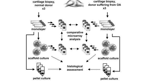
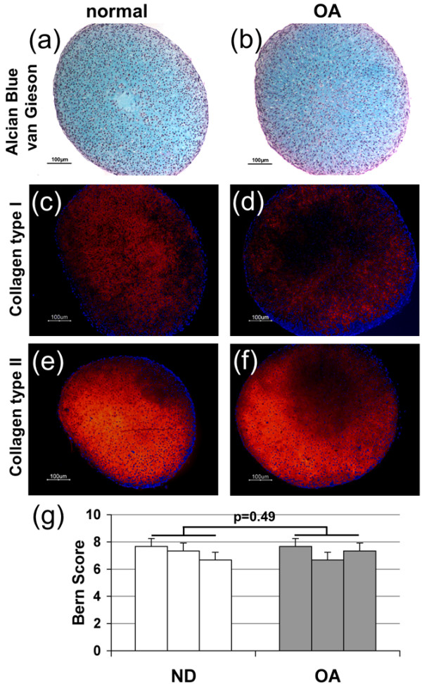
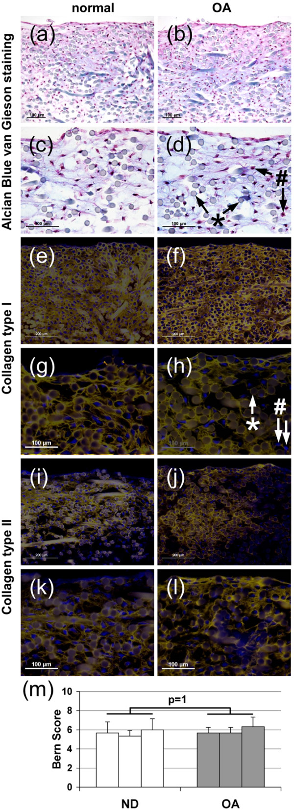
Methods: Articular chondrocytes from patients with OA undergoing total knee replacement (Mankin Score > 3, Ahlbäck Score > 2) and from patients undergoing ACT, here referred to as normal donors (ND), were isolated applying protocols used for ACT. Their chondrogenic differentiation potential was evaluated both in high-density pellet and scaffold (Hyaff-11) cultures by histological proteoglycan assessment (Bern Score) and immunohistochemistry for collagen types I and II. Chondrocytes cultured in monolayer and scaffolds were subjected to gene expression profiling using genome-wide oligonucleotide microarrays. Expression data were verified by using real-time PCR.

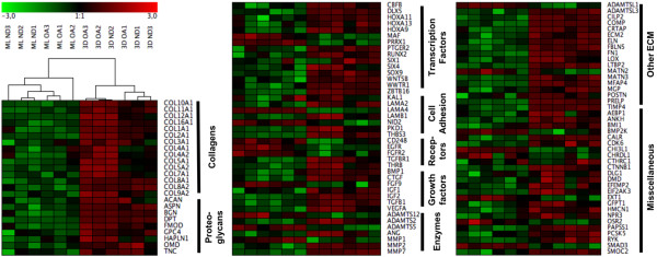
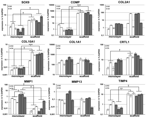
Results: Chondrocytes from ND and OA donors demonstrated accumulation of comparable amounts of cartilage matrix components, including sulphated proteoglycans and collagen types I and II. The mRNA expression of cartilage markers (ACAN, COL2A1, COMP, CRTL1, SOX9) and genes involved in matrix synthesis (BGN, CILP2, COL9A2, COL11A1, TIMP4) was highly induced in 3D cultures of chondrocytes from both donor groups. Genes associated with hypertrophic or OA cartilage (ALPL, COL1A1, COL3A1, COL10A1, MMP13, POSTN, PTH1R, RUNX2) were not significantly regulated between the two groups of donors. The expression of 661 genes, including COMP, FN1, and SOX9, was differentially regulated between OA and ND chondrocytes cultured in monolayer. During scaffold culture, the differences diminished between the OA and ND chondrocytes, and only 184 genes were differentially regulated.

Conclusions: Only few genes were differentially expressed between OA and ND chondrocytes in Hyaff-11 culture. The risk of differentiation into hypertrophic cartilage does not seem to be increased for OA chondrocytes. Our findings suggest that the chondrogenic capacity is not significantly affected by OA, and OA chondrocytes fulfill the requirements for matrix-associated ACT.


Figures

Figure 1
Figure 1
Schematic illustration of experimental setup. Articular chondrocytes from three patients with osteoarthritis and from three patients undergoing autologous chondrocyte transplantation (ACT) were isolated applying protocols used for ACT. After expansion in monolayer the chondrogenic differentiation potential was evaluated in high-density pellet and scaffold (Hyaff-11) cultures by histological assessment (Bern Score, immunohistochemistry for collagen types I and II). Chondrocytes cultured in monolayer and scaffolds were subjected to comparative gene expression analysis (genome-wide oligonucleotide microarrays, real-time PCR).
Figure 2
Figure 2
Histology of normal donor and osteoarthritic chondrocyte pellet cultures. Chondrogenic differentiation of chondrocytes obtained from (a, c, e) normal donors (ND) and (b, d, f) osteoarthritic (OA) articular cartilage using the high-density pellet culture system. (a, b) Alcian Blue van Gieson staining and immunohistochemical localization of (c, d) collagen type I and (e, f) type II. (g) Bern Score evaluating the differentiation grade of the cells. Three cultures per donor group.
Figure 3
Figure 3
Histology of osteoarthritic and normal chondrocyte scaffold culture. Chondrogenic differentiation of chondrocytes obtained from (a, c, e, g, i, k) normal and (b, d, f, h, j, l) osteoarthritic (OA) articular cartilage cultured in Hyaff-11 scaffolds. (a to d) Alcian Blue van Gieson staining, immunohistochemical localization of collagen (e to h) type I and (I to l) type II, with (g, h, k, l) higher magnification, and (m) Bern Score, * scaffold fibre, # cell nuclei. Three cultures per donor group.
Figure 4
Figure 4
Hierarchical cluster analysis of chondrocytes from osteoarthritic and normal donors cultured in monolayer and Hyaff-11 scaffolds. Genes that were differentially expressed between normal donors (ND) chondrocytes cultured in monolayer (ML) and scaffold (3D) cultures, functionally filtered by their association with skeletal development and extracellular matrix (ECM) formation, were used to assess chondrogenic capacity of chondrocytes from osteoarthritic (OA) patients. Green bars depict a repressed and red bars an induced expression of genes normalized to the mean. The clustering gave two main groups classified as monolayer chondrocytes and scaffold-cultured chondrocytes. The separate OA monolayer cluster clearly indicated a differential expression pattern between OA and ND chondrocytes. In scaffold cultures on the other hand, no OA-related cluster separation was observed demonstrating a loss of differences between OA and ND chondrocytes during scaffold culture.
Figure 5
Figure 5
Real-time PCR verification of results from the microarray analysis. The expression was calculated as percentage of the expression of the housekeeping gene glyceraldehyde-3-phosphate dehydrogenase (GAPDH). The mean of each technical triplicate is plotted and the error bars represent standard deviation. * P < 0.05; ** P < 0.01; *** P < 0.001.

References

    1. Alford JW, Cole BJ. Cartilage restoration, part 1: basic science, historical perspective, patient evaluation, and treatment options. The American journal of sports medicine. 2005;33:295–306. doi: 10.1177/0363546504273510. - DOI - PubMed
    1. Brittberg M, Lindahl A, Nilsson A, Ohlsson C, Isaksson O, Peterson L. Treatment of deep cartilage defects in the knee with autologous chondrocyte transplantation. The New England journal of medicine. 1994;331:889–895. doi: 10.1056/NEJM199410063311401. - DOI - PubMed
    1. Behrens P, Bitter T, Kurz B, Russlies M. Matrix-associated autologous chondrocyte transplantation/implantation (MACT/MACI)--5-year follow-up. The Knee. 2006;13:194–202. doi: 10.1016/j.knee.2006.02.012. - DOI - PubMed
    1. Marcacci M, Berruto M, Brocchetta D, Delcogliano A, Ghinelli D, Gobbi A, Kon E, Pederzini L, Rosa D, Sacchetti GL, Stefani G, Zanasi S. Articular cartilage engineering with Hyalograft C: 3-year clinical results. Clinical orthopaedics and related research. 2005. pp. 96–105. - DOI - PubMed
    1. Ossendorf C, Kaps C, Kreuz PC, Burmester GR, Sittinger M, Erggelet C. Treatment of posttraumatic and focal osteoarthritic cartilage defects of the knee with autologous polymer-based three-dimensional chondrocyte grafts: 2-year clinical results. Arthritis Res Ther. 2007;9:R41. doi: 10.1186/ar2180. - DOI - PMC - PubMed

Publication types