

A current view of brain renin-angiotensin system: Is the (pro)renin receptor the missing link?
- PMID: 19723538
- PMCID: PMC2815255
- DOI: 10.1016/j.pharmthera.2009.07.007
A current view of brain renin-angiotensin system: Is the (pro)renin receptor the missing link?
Abstract
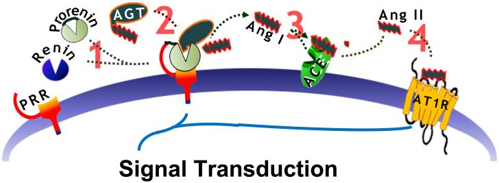
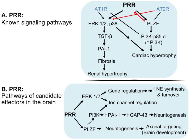
The renin-angiotensin system (RAS) plays a central role in the brain to regulate blood pressure (BP). This role includes the modulation of sympathetic nerve activity (SNA) that regulates vascular tone; the regulation of secretion of neurohormones that have a critical role in electrolyte as well as fluid homeostasis; and by influencing behavioral processes to increase salt and water intake. Based on decades of research it is clear that angiotensin II (Ang II), the major bioactive product of the RAS, mediates these actions largely via its Ang II type 1 receptor (AT1R), located within hypothalamic and brainstem control centers. However, the mechanisms of brain RAS function have been questioned, due in large part to low expression levels of the rate limiting enzyme renin within the central nervous system. Tissue localized RAS has been observed in heart, kidney tubules and vascular cells. Studies have also given rise to the hypothesis for localized RAS function within the brain, so that Ang II can act in a paracrine manner to influence neuronal activity. The recently discovered (pro)renin receptor (PRR) may be key in this mechanism as it serves to sequester renin and prorenin for localized RAS activity. Thus, the PRR can potentially mitigate the low levels of renin expression in the brain to propagate Ang II action. In this review we examine the regulation, expression and functional properties of the various RAS components in the brain with particular focus on the different roles that PRR may have in BP regulation and hypertension.
Figures
References
-
- Allen AM. Blockade of angiotensin AT1-receptors in the rostral ventrolateral medulla of spontaneously hypertensive rats reduces blood pressure and sympathetic nerve discharge. J Renin Angiotensin Aldosterone Syst. 2001;2:s120–124. - PubMed
-
- Allen AM, et al. Expression of constitutively active angiotensin receptors in the rostral ventrolateral medulla increases blood pressure. Hypertension. 2006;47:1054–1061. - PubMed
-
- Allen AM, MacGregor DP, McKinley MJ, Mendelsohn FA. Angiotensin II receptors in the human brain. Regul Pept. 1999;79:1–7. - PubMed
-
- Averill DB, Diz DI. Angiotensin peptides and baroreflex control of sympathetic outflow: pathways and mechanisms of the medulla oblongata. Brain Res Bull. 2000;51:119–128. - PubMed
Publication types
MeSH terms
Substances
Grants and funding
LinkOut - more resources
Full Text Sources
Other Literature Sources
Medical
Research Materials
Miscellaneous
